Article Text

Abstract
Background Structural pathology may be present in joints without radiographic evidence of osteoarthritis (OA). Ultrasound is a sensitive tool for early detection of osteophytes. Our aim was to explore whether ultrasound-detected osteophytes (in radiographically and clinically normal finger joints) predicted the development of radiographic and clinical hand OA 5 years later.
Methods We included finger joints without radiographic OA (Kellgren-Lawrence grade (KLG)=0; n=301) or no clinical bony enlargements (n=717) at baseline and examined whether ultrasound-detected osteophytes predicted incident radiographic OA (KLG ≥1, osteophytes or joint space narrowing (JSN)) or incident clinical bony enlargement (dependent variables) in the same joints 5 years later. We applied logistic regression with generalised estimating equations adjusted for age, sex, body mass index and follow-up time.
Results Ultrasound demonstrated osteophytes in 86/301 (28.6%) joints without radiographic OA and 392/717 (54.7%) joints without clinical bony enlargement. These osteophytes were confirmed in the majority of joints where MRI assessment was available. Significant associations were found between ultrasound-detected osteophytes and development of both radiographic OA (OR=4.1, 95% CI 2.0 to 8.1) and clinical bony enlargement (OR=3.5, 95% CI 2.4 to 5.1) and also incident radiographic osteophytes (OR=4.2, 95% CI 2.1 to 8.5) and JSN (OR=5.3, 95% CI 2.1 to 13.4).
Conclusion Ultrasound-detected osteophytes predicted incident radiographic and clinical hand OA 5 years later. These results support the use of ultrasound for early detection of OA.
- hand osteoarthritis
- ultrasonography
- epidemiology
This is an Open Access article distributed in accordance with the Creative Commons Attribution Non Commercial (CC BY-NC 4.0) license, which permits others to distribute, remix, adapt, build upon this work non-commercially, and license their derivative works on different terms, provided the original work is properly cited and the use is non-commercial. See: http://creativecommons.org/licenses/by-nc/4.0/
Statistics from Altmetric.com
Key messages
What is already known about this subject?
Osteophyte formation is considered a key feature of osteoarthritis (OA) leading to pain and loss of function.
Cross-sectional studies in both knee and hand OA have demonstrated that ultrasound is more sensitive than conventional radiography in detecting osteophytes, but little is known about the implication of osteophytes detected by ultrasound before OA becomes apparent on radiographs or clinical examination.
What does this study add?
Our longitudinal study demonstrated that ultrasound-detected osteophytes (in joints assessed as normal on radiographs and clinical examination) predicted future development of radiographic and clinical osteoarthritic features in the same finger joints.
How might this impact on clinical practice?
Taken together with a series of other studies, our data suggest that sensitive imaging modalities such as ultrasound (or MRI) should be applied when an early hand OA diagnosis is warranted.
Introduction
In the field of inflammatory rheumatic disorders, growing evidence has made it clear that early and targeted treatment significantly improves prognosis.1 Similar strategies may be applied to patients with osteoarthritis (OA) if disease-modifying OA drugs (DMOADs) become available.2 3 Conventional radiography remains the cornerstone in obtaining an image-based OA diagnosis.4 However, previous studies on knee OA have shown that cartilage degradation is well established and often substantial by the time radiographic changes are identified.5 6 Hence, we need instruments to identify OA at an earlier stage.
Osteophyte formation is considered a key feature of OA leading to pain and loss of function.7 Knee OA studies suggest that osteophytes represent a more reliable indicator of early disease than joint space narrowing (JSN).7 Cross-sectional studies in both knee and hand OA have demonstrated that ultrasound is more sensitive than conventional radiography in detecting osteophytes,8–10 whereas few studies have explored the predictive value of osteophytes on hand OA progression. In 74 patients with hand OA, MRI-defined osteophytes predicted the development of erosions 5 years later, but no statistically significant associations were found for radiographic progression according to Kellgren-Lawrence grade (KLG) or JSN.11
To our knowledge, no previous study has explored whether ultrasound-detected osteophytes predict future development of radiographic OA. The present objective was to determine whether ultrasound-detected osteophytes in finger joints without radiographic or clinical OA at baseline could predict the development of radiographic or clinical hand OA 5 years later.
Methods
Patients
Participants in the Oslo Hand OA cohort were recruited from the rheumatology outpatient clinic at Diakonhjemmet Hospital (Oslo, Norway) in 2001–2003 (n=209) with follow-up examinations in 2008–2009 (n=128) and 2013 (n=87).12 13 We included men/women (50–70 years) with hand OA and no diagnosis of systemic inflammatory rheumatic disease.
In the current analyses, we used data from 2008 to 2009 (hereafter referred to as ‘baseline’) and 2013 (hereafter referred to as ‘follow-up’) due to no ultrasound examination in 2001–2003. Of the 87 patients who were examined in 2013, 78 participants had available ultrasound examination at baseline and conventional radiography as well as clinical examination at baseline and follow-up. The 78 participants who were included in analyses and the nine patients who were excluded due to missing data had similar gender distribution (p=0.35), mean age (p=0.59) or KLG sum score at follow-up (p=0.76).
The regional ethics committee approved the study, and all participants gave their written informed consent.
Ultrasound
Sonographic examination of hands was performed at baseline using the same ultrasound machine (Siemens Medical Solutions, Excellence version, Mountain View, California, USA) with fixed settings and a 5–13 MHz linear array transducer. Blinded to clinical and other imaging results, two sonographers (AM and HBH) performed the assessments together and reached consensus on each scoring. The scanning protocol and ‘very good’ reliability have been described previously.8 The sonographers scored osteophytes in each finger joint according to a semiquantitative scoring system (grade 0–3).8 In the current analyses, we included the n=20 joints most likely to develop OA: the bilateral first carpometacarpal (CMC-1) (longitudinal palmolateral scan), the thumb interphalangeal (IP-1) and the second to fifth proximal interphalangeal and distal interphalangeal (PIP and DIP, respectively) joints (longitudinal dorsal scan from the radial to the ulnar side).
Conventional radiography
Bilateral hand radiographs (posteroanterior view) were obtained at baseline and follow-up. Blinded to clinical and ultrasound findings, one reader (IKH) scored the paired images (20 joints, same as ultrasound) with known time sequence for OA according to KLG (grade 0–4), as well as osteophytes (grade 0–3) and JSN (grade 0–3) according to the Osteoarthritis Research Society International atlas.14 15Intrareader reliability for radiographic status and change scores were ‘good’ to ‘very good’.16
Incident radiographic OA at follow-up was defined as an increase in KLG from 0 to 1–4, and incident radiographic osteophytes and incident JSN both corresponded with an increase from grade 0 to grades 1–3.
Magnetic resonance imaging
With a 1.0 T extremity MRI unit (ONI; GE Healthcare, Waukesha, Wisconsin, USA), presence of osteophytes was examined in second to fifth DIP and PIP joints of the dominant hand in 73 participants. The acquisition, scoring and good reliability have previously been described.8
Clinical examination
Blinded to imaging results, one experienced rheumatologist (BSC) examined the above-mentioned 20 joints for bony enlargements on palpation (‘absent' or ‘present') at baseline and follow-up.
Incident clinical OA at follow-up was defined as development of bony enlargement (from absent to present).
Statistics
Data are presented as mean (SD) values. Independent samples t-test, Mann-Whitney U test and Fisher’s exact test were applied as appropriate to compare variables.
In longitudinal analyses at the joint level, we applied logistic regression with generalised estimating equations (exchangeable correlation matrix), presented as OR with 95% CI. We selected joints with KLG=0 or no clinical bony enlargements at baseline and examined whether ultrasound-detected osteophytes (independent variable) could predict incident radiographic OA (KLG ≥1, osteophytes or JSN) or clinical bony enlargement (dependent variables) in the same joint at follow-up. Joints without sonographic osteophytes served as reference. Analyses were adjusted for age, sex, body mass index at baseline and follow-up time. Missing joints were due to unilateral radiographs (n=3 participants), trapezectomy (12 joints), fixation (6 joints) and unrecorded ultrasound findings (4 joints).
Statistical analyses were performed using IBM SPSS Statistics V.24.0 (IBM, Armonk, New York, USA).
Results
Mean (SD) age at baseline was 67.8 (5.2) years and 91% were women. Mean (SD) follow-up time was 4.7 (0.4) years.
At baseline, 1508 joints had available radiographic and ultrasound examinations, of which 301 (20.0%) joints were assessed normal on conventional radiographs (KLG=0). In these radiographically normal joints, ultrasound demonstrated osteophytes in 28.6% joints (table 1a), and highest discordance was found in DIP joints with sonographic osteophytes present in 53.2% joints. The majority of these osteophytes (79.1%) were small (grade=1). In joints with radiographic ‘doubtful OA’ (KLG=1, n=186) and ‘definite OA’ (KLG=2–4, n=1021), sonographic osteophytes were found in 62.9% and 90.7% joints, respectively (table 1b and c). Similarly, in 717/1508 joints without clinical bony enlargement at baseline, sonographic osteophytes were demonstrated in 54.7% joints (table 1d).
Baseline data: ultrasound-detected osteophytes in joints assessed as (A) normal, (B) doubtful OA or (C) definite OA on radiographs, as well as in (D) clinical normal joints
In a limited number of joints with available MRI, we were able to confirm the presence of these baseline sonographic osteophytes (ie, positive predictive value) in 86.5% (n=32/37), 90.2% (n=37/41) and 97.0% (n=356/367) of the joints assessed as KL=0, KL=1 and KL=2–4 by radiographs, respectively, and similarly in 91.6% (n=120/131) and 97.1% (n=305/314) joints without or with clinical bony enlargement, respectively.
In longitudinal analyses, incident radiographic and clinical OA occurred during follow-up in 46.5% and 60.5% of joints with baseline sonographic osteophytes. Analyses on joint level demonstrated that ultrasound-detected osteophytes (in radiographically and clinically normal finger joints at baseline) significantly predicted development of radiographic OA according to KLG and clinical bony enlargement during follow-up (table 2a and c and figure 1) and also a development of radiographic osteophytes and JSN (table 2a). Sonographic osteophytes predicted radiographic OA even stronger when incident radiographic OA was defined as KLG ≥2 at follow-up (table 2b). Analyses including non-steroidal anti-inflammatory drugs or oral glucocorticoids did not change our results. Statistically significant associations between baseline ultrasound-detected osteophytes and incident radiographic OA was found in PIP and DIP joints but not CMC-1 and IP-1, in which the association was statistically non-significant due to few joints with incident radiographic OA (online supplementary table). Significant associations were demonstrated for sonographic osteophytes and incident clinical bony enlargement in all joint groups (online supplementary table).
Supplementary file 1
Associations between ultrasound-detected osteophytes at baseline (independent variables) and incident radiographic or clinical OA at follow-up (dependent variables in separate models)

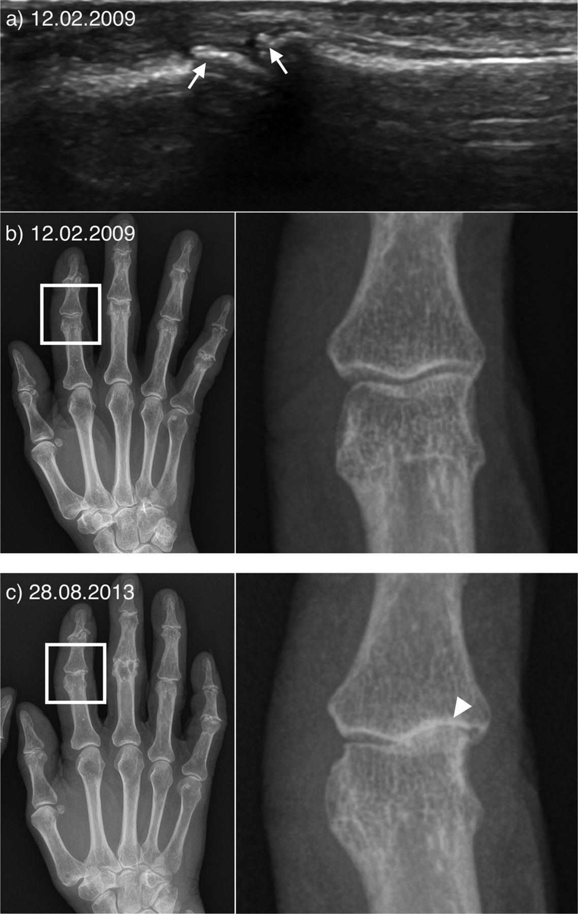
Ultrasound examination and conventional radiography of the second proximal interphalangeal joint at baseline (2009) and follow-up (2013). Ultrasound (A) showed small osteophytes at the proximal and distal joint surface (arrows), while concurrent radiographs (B) was assessed as normal (Kellgren-Lawrence grade=0). At followup (C), the same joint had progressed to radiographic OA (arrowhead), with development of joint space narrowing and subchondral sclerosis (arrowhead) as well as malalignment.
In a separate analysis, we compared progression in joints with baseline radiographic KL grade=0 and KL grade=1. Interestingly, 35% (n=65/186) of joints categorised as ‘doubtful OA’ (ie, KL grade=1) at baseline went on to develop true hand OA (KL grades 2–4) after 5 years compared with only 9% (27/301) of joints with no radiographic sign of disease at baseline (p<0.001). When further stratifying for presence of baseline sonographic osteophytes, 41.0% versus 24.6% of the joints developed true OA (p=0.006).
Discussion
In this longitudinal observational hand OA study, ultrasound-detected osteophytes were frequently found in finger joints with no radiographic OA and no clinical bony enlargements. Furthermore, the presence of ultrasound-detected osteophytes strongly predicted incident radiographic and clinical OA 5 years later at joint level, supporting that ultrasound is more sensitive than radiography and clinical examination to detect early hand OA.
Osteophytes, mostly small, were found by ultrasound in 29% and 55% of joints assessed as normal on radiographs and clinical examination (table 1), respectively. Our findings are in line with those of Keen and colleagues, who reported sonographic osteophytes in 24% finger joints that were normal on radiographs.17 In knee OA, Guermazi et al found osteophytes by MRI in as much as 74% of knee joints with no radiographic features of OA,18 but prevalence was only 14% when stringent definitions of MRI osteophytes were applied. With the radiographic posteroanterior view of the hands, small osteophytes may go unnoticed on radiographs, especially when present on the dorsal or palmar aspects of the joints. Our findings highlight the limitation of conventional radiography to diagnose a large number of finger joints with small osteophytes. This was especially true in DIP joints, where sonographic osteophytes were found in half of the joints assessed as normal on radiographs, and although MRI assessment was available in a limited number of joints and patients, the high positive predictive values indicate that (even small) osteophytes detected by ultrasound in our cohort were, in fact, true osteophytes.
Furthermore, with longitudinal data, we had the opportunity to explore the importance of these early-detected sonographic osteophytes, which were shown to strongly predict development of radiographic and clinical OA at follow-up (table 2). Most predictive studies have been performed on patients with knee OA. Saunders et al found that both JSN and osteophytes act as independent predictors of cartilage volume loss over a 2-year period in a large cohort of randomly selected older adults.19 With data from the Osteoarthritis Initiative study, Roemer and colleagues recently evaluated MRI of knee OA patients at multiple time points prior to radiographic disease onset6 and found that the number of features (or involved structures) were more important than any single feature.6 Ultrasonography enables examination of several aspects of the joint, both bony changes such as osteophytes and erosions, and soft tissue changes such as synovial effusion, hypertrophy and vascularisation. MRI has a limited role in everyday clinical practice due to costs and availability, whereas our results support ultrasound as a complementary imaging tool along with radiography for more accurate diagnostics of hand OA.
The relation between osteophytes and radiographic progression is complex and multifactorial. Several structures of the joint may at some point individually or together drive the disease.6 In animal models, osteophytes develop at sites adjacent to cartilage loss.20 While OA knees with large osteophytes are more likely to progress than knees without osteophytes, it is assumed that the strong relation between osteophytes and malalignment, in part, explains the progression.21 It is further suggested that osteophytes do not have any direct role on disease progression but serve as markers of the location and severity of other pathologic processes.21 Thus, the importance of the present results is that ultrasound-detected osteophytes may be markers of early joint changes (and not having a causal effect on OA progression).
Still, conventional radiography will remain the cornerstone in obtaining an image-based OA diagnosis in our daily clinical practice. However, the most accepted scoring system, the KL scale, is criticised over its inconsistencies of grade 1. Do these ‘doubtful changes’ of osteophytes or JSN represent normal joints or early OA? In our cohort, we found progression to ‘true OA’ in significantly more joints with baseline KL grade=1 than KL grade=0. Similarly, a 10-year study found 62% of women having small radiographic tibiofemoral osteophytes at baseline to develop true osteophytic knee OA during follow-up compared with only 22% of controls with no signs of disease at baseline.22 We support the conclusion by Hart et al: so-called ‘doubtful’ osteophytes appear to be ‘real’ and cannot be ignored or classified as normal.22
Our study is limited by the high age of patients with mostly extensive OA at baseline. However, although patients already have established disease, they still had joints with no OA. A similar study on patients not fulfilling the criteria for hand OA at baseline is of interest. It is also uncertain whether our results can be generalised to other OA joints. Third, radiographs were scored in known time sequence, which may lead to overestimation of progression. However, blinding may increase the error rate, and unblinding is the recommended approach to serial images.23 Finally, we do not have reliability for the bony enlargement examination.
Conclusion
Our study provides evidence confirming that ultrasound may identify finger osteophytes at an earlier stage than conventional radiographs and clinical examinations. Early identification of preradiographic OA may be especially important in the event of a future DMOAD.
Acknowledgments
We would like to thank the participants of the Oslo HOA cohort and the staff at the Department of Rheumatology at Diakonhjemmet Hospital (Oslo, Norway) for helping us to perform this study.
References
Footnotes
Contributors Data collection: AM, BS-C, HBH and IKH. Study design: AM and IKH. Analyses, interpretation of data and drafting the paper: AM. Critical revision and final approval: AM, BS-C, HBH, IKH and TKK.
Funding ‘The Norwegian ExtraFoundation for Health and Rehabilitation’ through EXTRA funds has financed the PhD position of AM.
Competing interests None declared.
Patient consent Obtained.
Ethics approval Regional Ethical Committee (Norway).
Provenance and peer review Not commissioned; externally peer reviewed.
Data sharing statement Data available upon written request.