Article Text

Abstract
This paper presents an atlas for the Outcome Measures in Rheumatology Clinical Trials (OMERACT) thumb base osteoarthritis MRI scoring system (TOMS). The atlas includes reference images of each grade of each feature that is assessed in TOMS (synovitis grade 0–3, subchondral bone defects grade 0–3, osteophytes grade 0–3, cartilage assessment grade 0–3, subluxation and bone marrow lesions grade 0–3) in the first carpometacarpal and scapho-trapezio-trapezoid joint. The presented reference images can be used to guide scoring of thumb base MRIs in patients with hand osteoarthritis according to the OMERACT TOMS.
- osteoarthritis
- magnetic resonance imaging
- hand osteoarthritis
This is an Open Access article distributed in accordance with the Creative Commons Attribution Non Commercial (CC BY-NC 4.0) license, which permits others to distribute, remix, adapt, build upon this work non-commercially, and license their derivative works on different terms, provided the original work is properly cited and the use is non-commercial. See: http://creativecommons.org/licenses/by-nc/4.0/
Statistics from Altmetric.com
Hand OA affects the interphalangeal and thumb base joints, including the CMC-1 and STT joints. Much is still unknown about the pathophysiology of thumb base OA. Although MRI studies have led to more insights in interphalangeal OA, thumb base MRI studies are still lacking. To facilitate this, recently the first MRI scoring system for thumb base OA was developed by the OMERACT MRI Working Group, the TOMS.1
Representative examples of each grade of the different features that are assessed in the TOMS are presented (see table 1 for definitions and scaling of each feature). Images from patients with hand OA were obtained from the Hand Osteoarthritis in Secondary Care (HOSTAS) study at Leiden University Medical Center (Leiden, The Netherlands). Images were acquired on a 1.5 T extremity MRI unit (ONI, GE, Wisconsin, USA). Examples of synovitis evaluated on contrast-enhanced images were obtained from patients with hand OA from the Nor-Hand study at Diakonhjemmet Hospital (Oslo, Norway) and were acquired on a 1.5 T MRI unit (Siemens Aera, Germany) after administration of gadolinium contrast. Example images were selected by a single reader with experience in using the TOMS and subsequently approved by three experienced radiologists (of which one is also experienced in using the score).
Definitions and scaling of features in proposed OMERACT TOMS
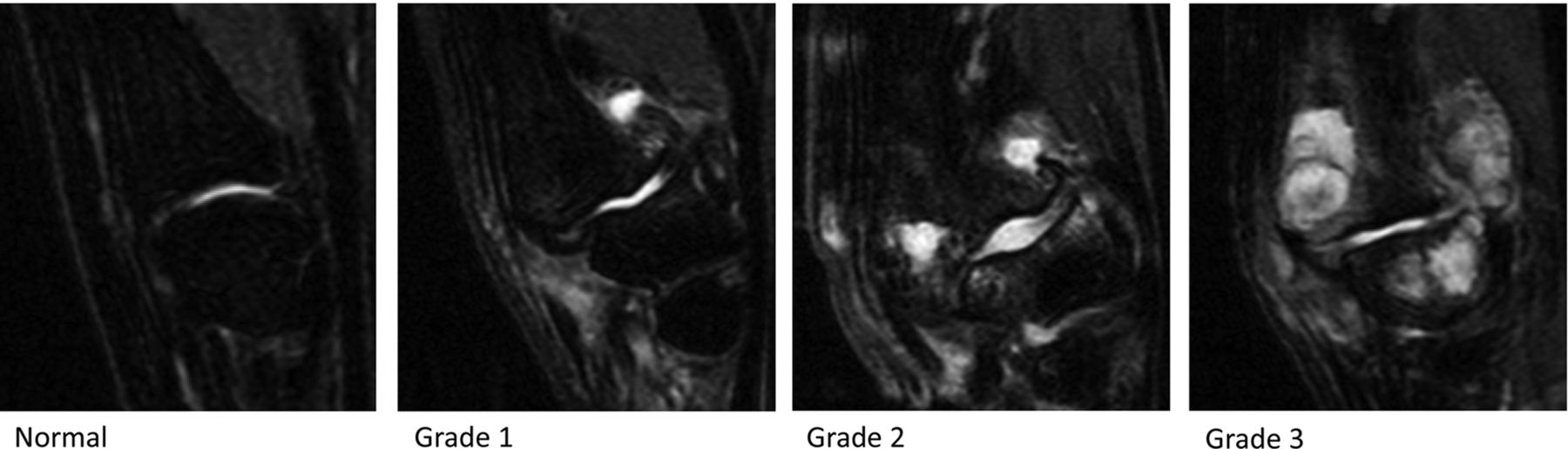
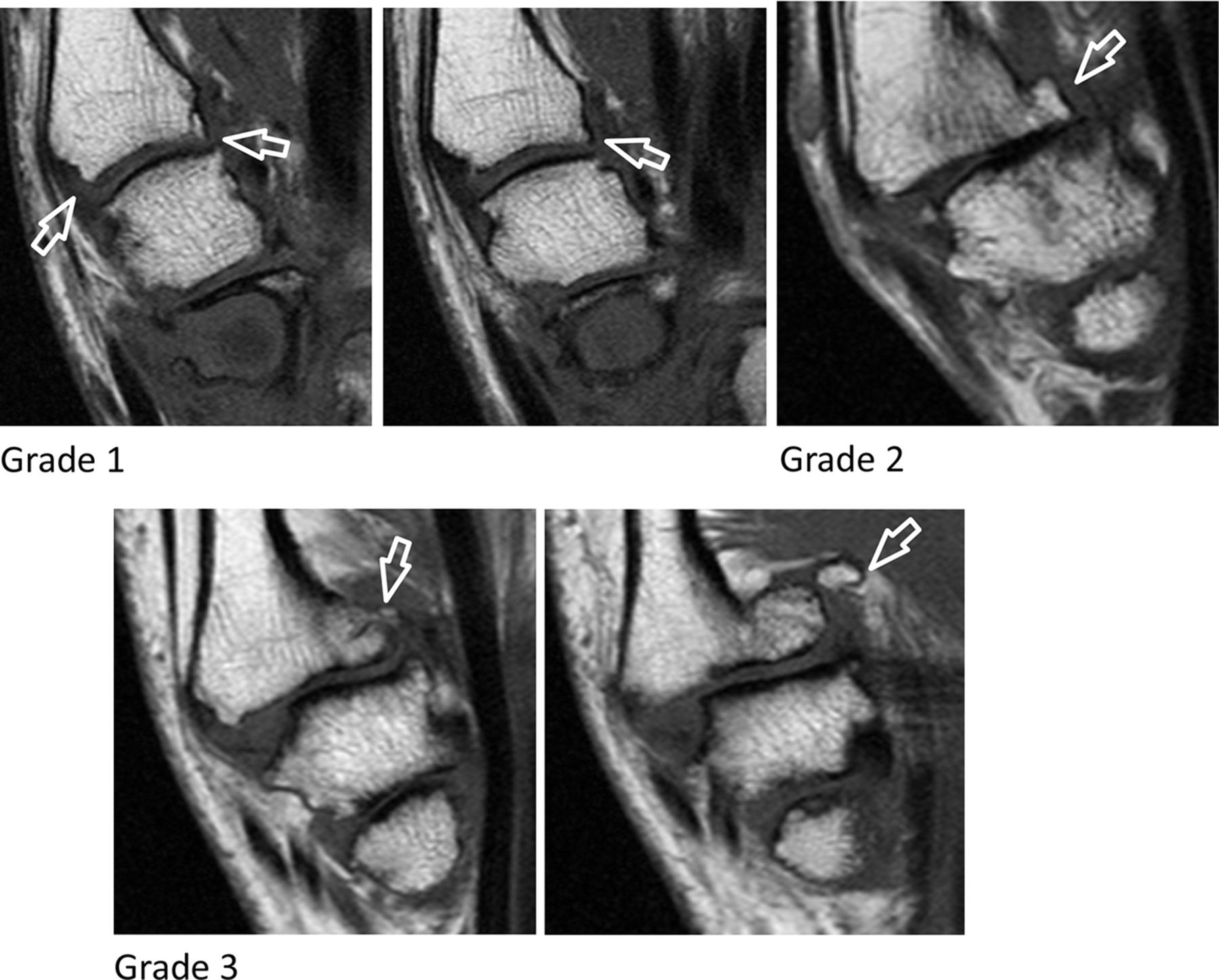
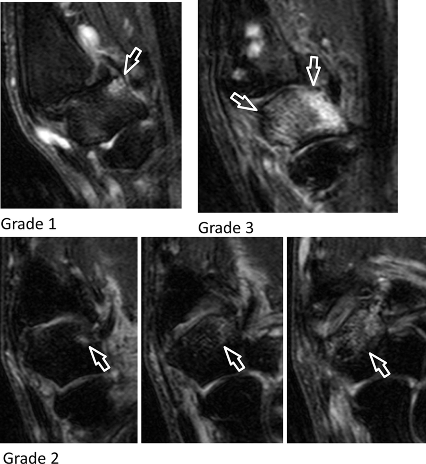
Synovitis first carpometacarpal (T1 weighted-fat saturated postcontrast images).

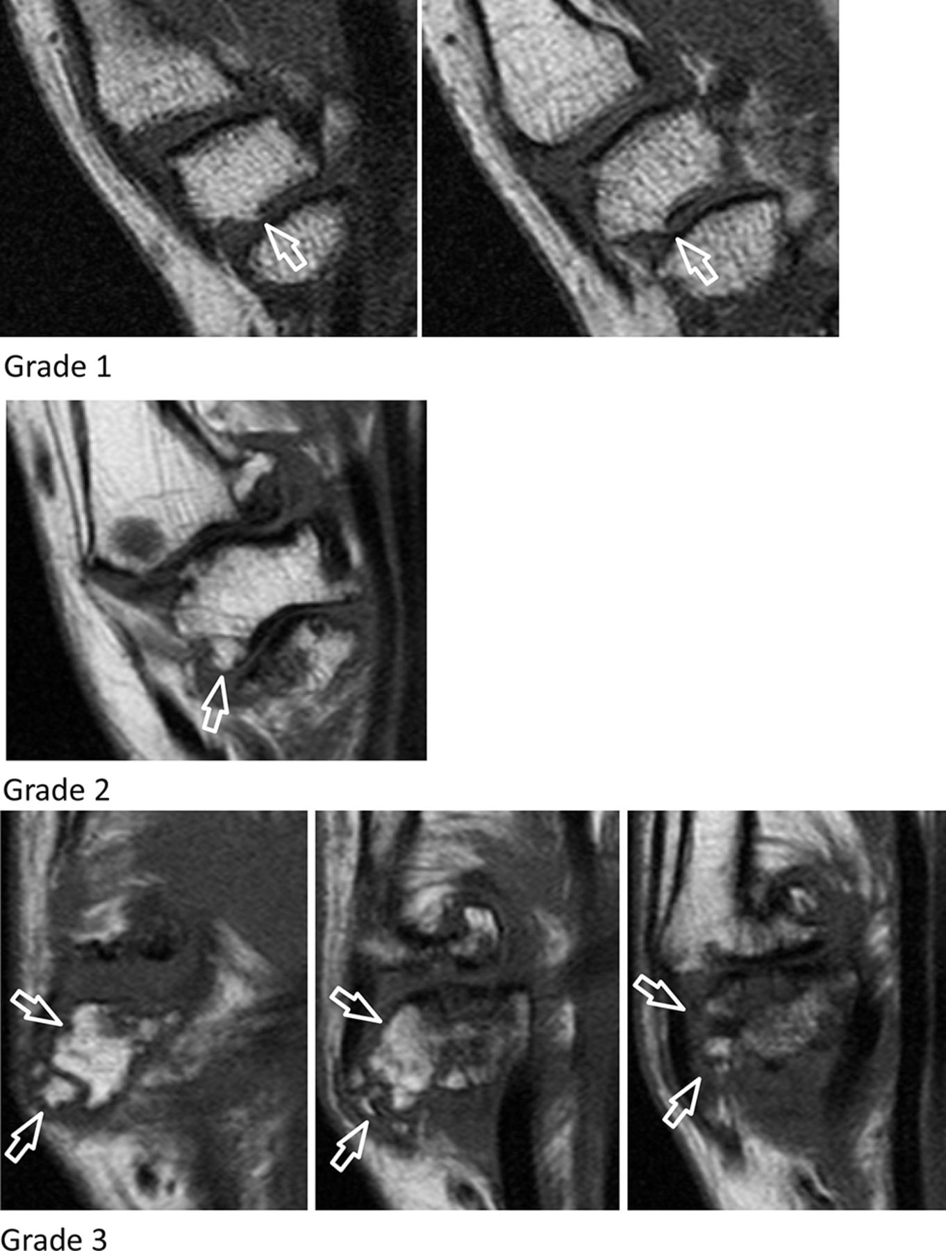
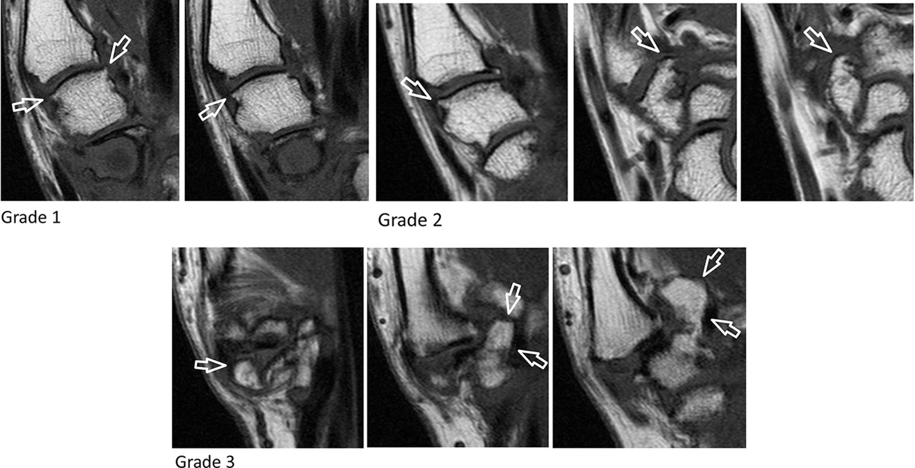
Synovitis first carpometacarpal (T2 weighted-fat saturated images).

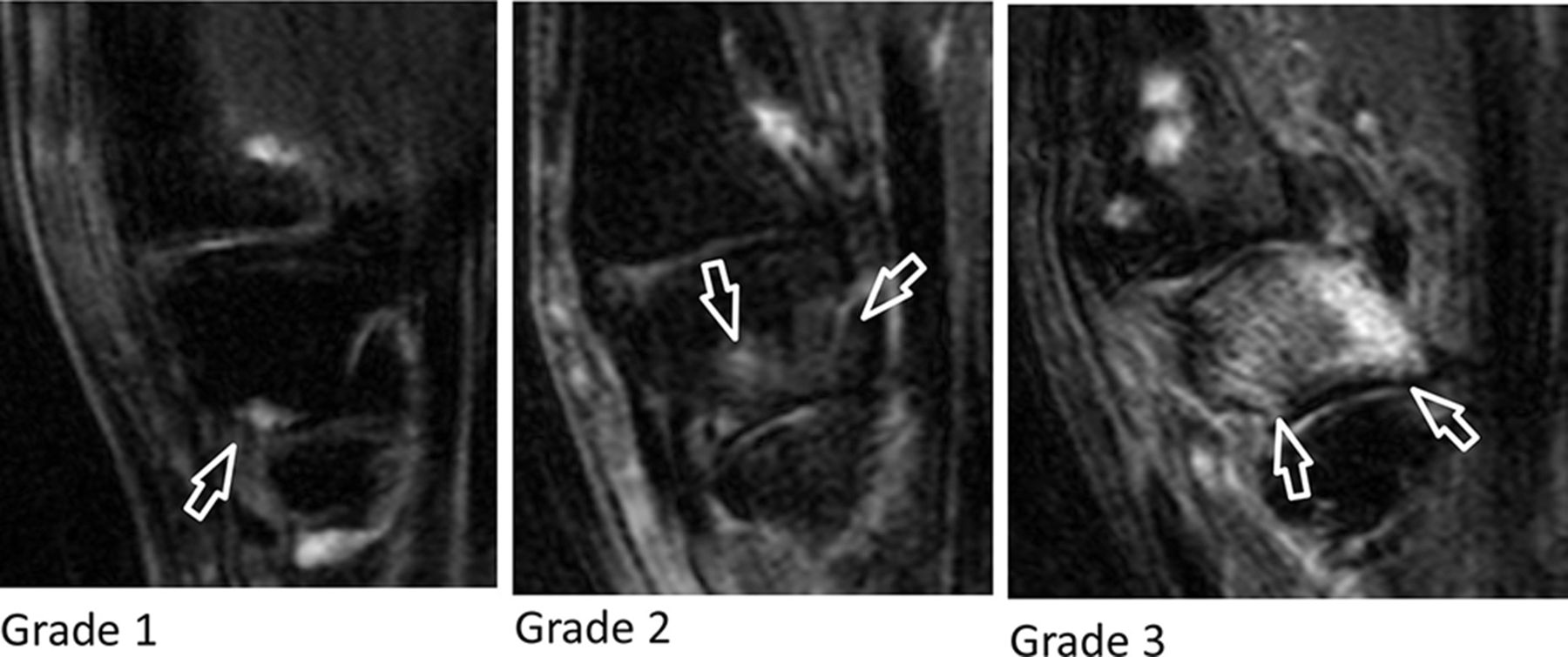
Synovitis scapho-trapezio-trapezoid (T2 weighted-fat saturated images).
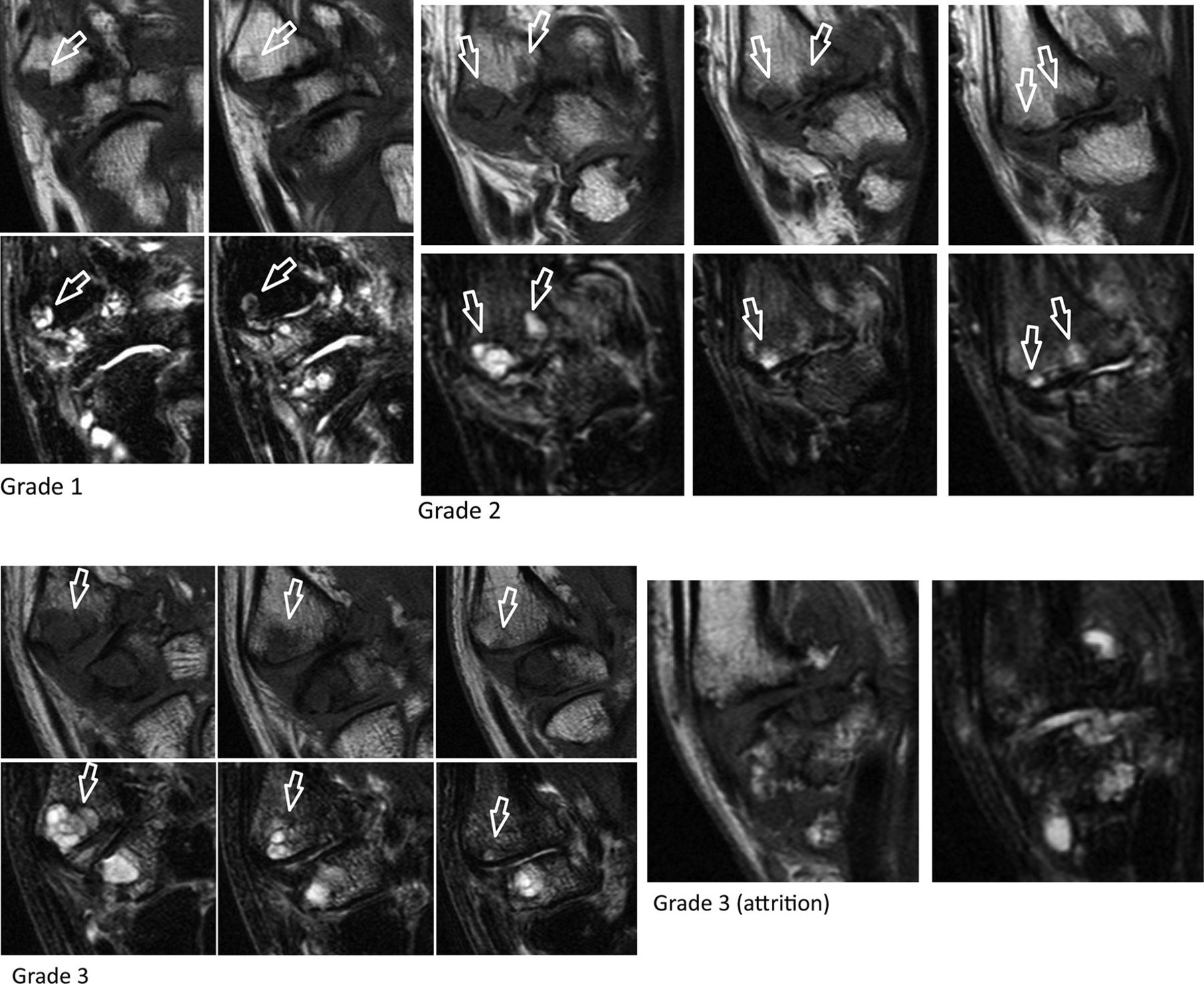
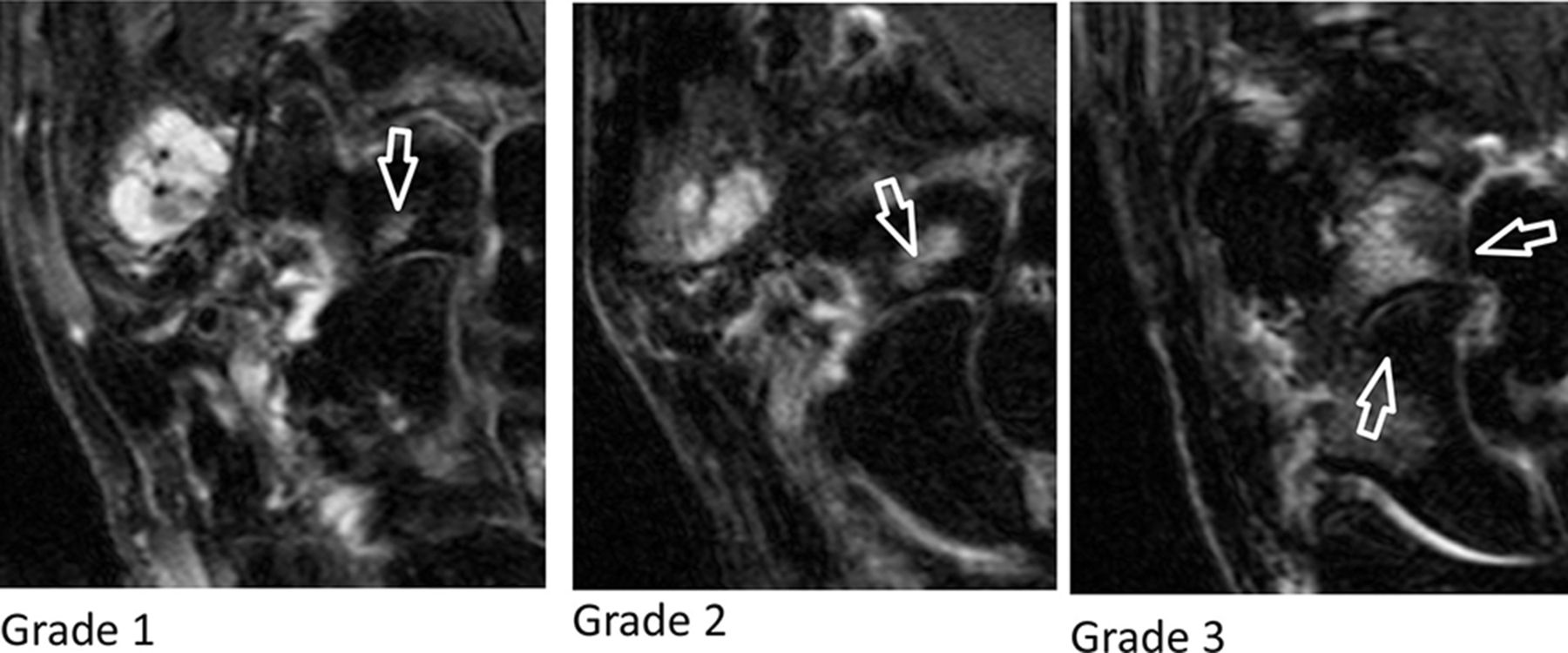
Subchondral bone defects first carpometacarpal: proximal first metacarpal (T1 weighted and T2 weighted-fat saturated images).
Subchondral bone defects first carpometacarpal: distal trapezium (T1 weighted and T2 weighted-fat saturated images).

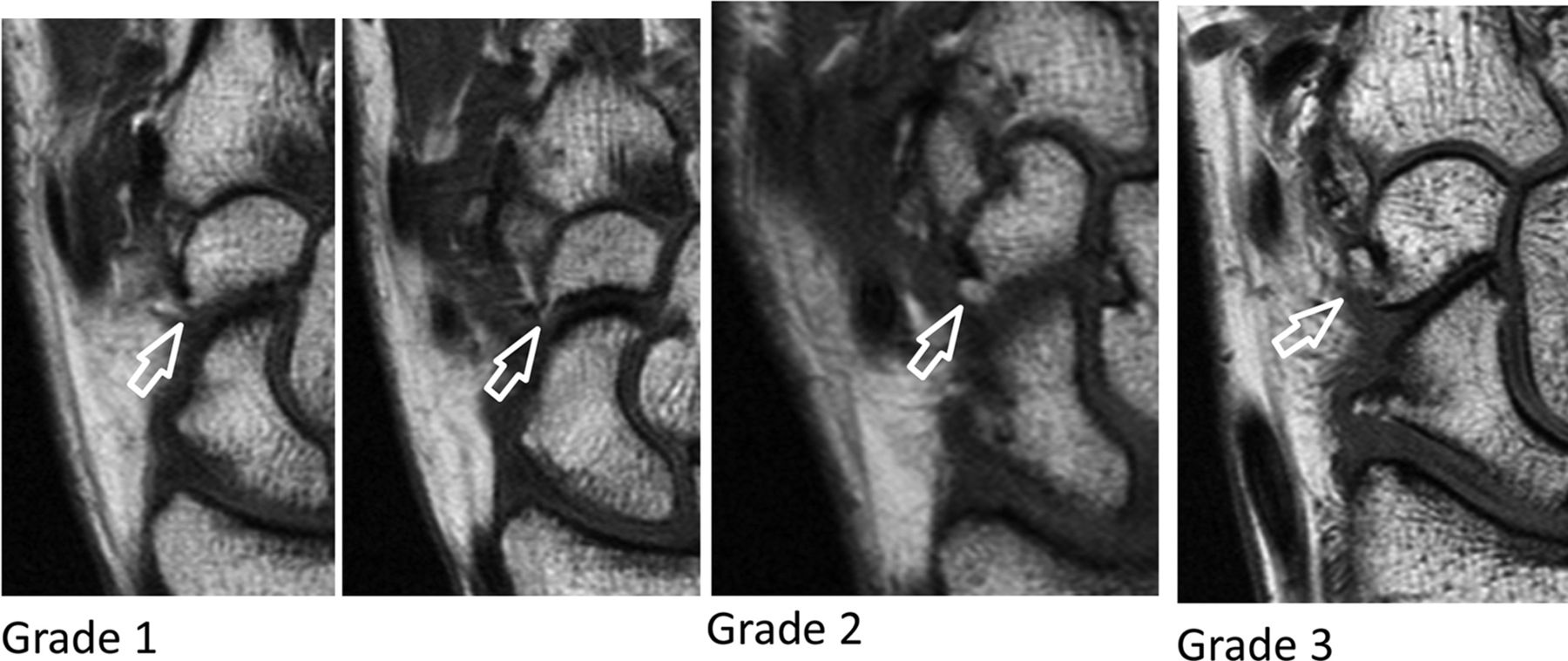
Subchondral bone defects scapho-trapezio-trapezoid: proximal trapezium (T1 weighted and T2 weighted-fat saturated images).

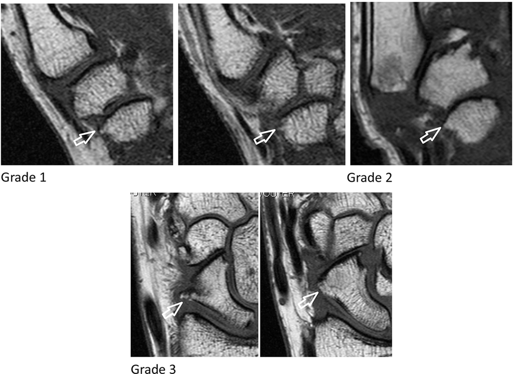
Subchondral bone defects scapho-trapezio-trapezoid: proximal trapezoid (T1 weighted and T2 weighted-fat saturated images).
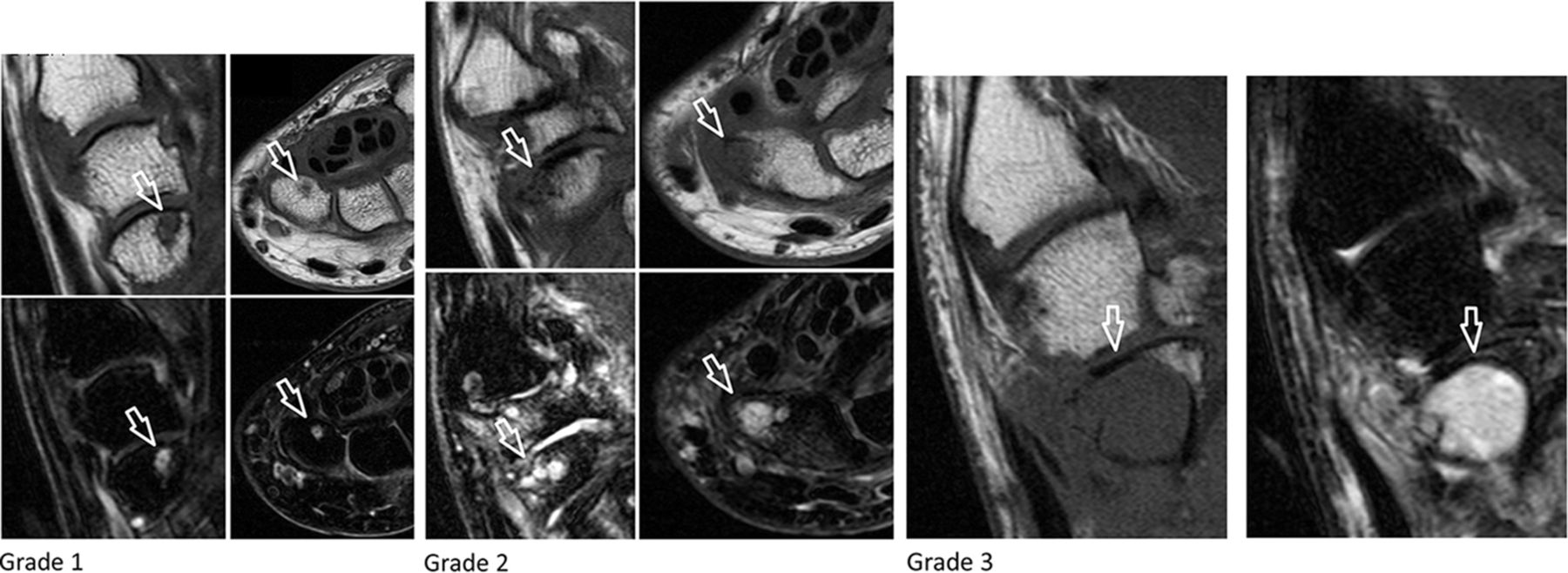
Subchondral bone defects scapho-trapezio-trapezoid: distal scaphoid (T1 weighted and T2 weighted-fat saturated images).

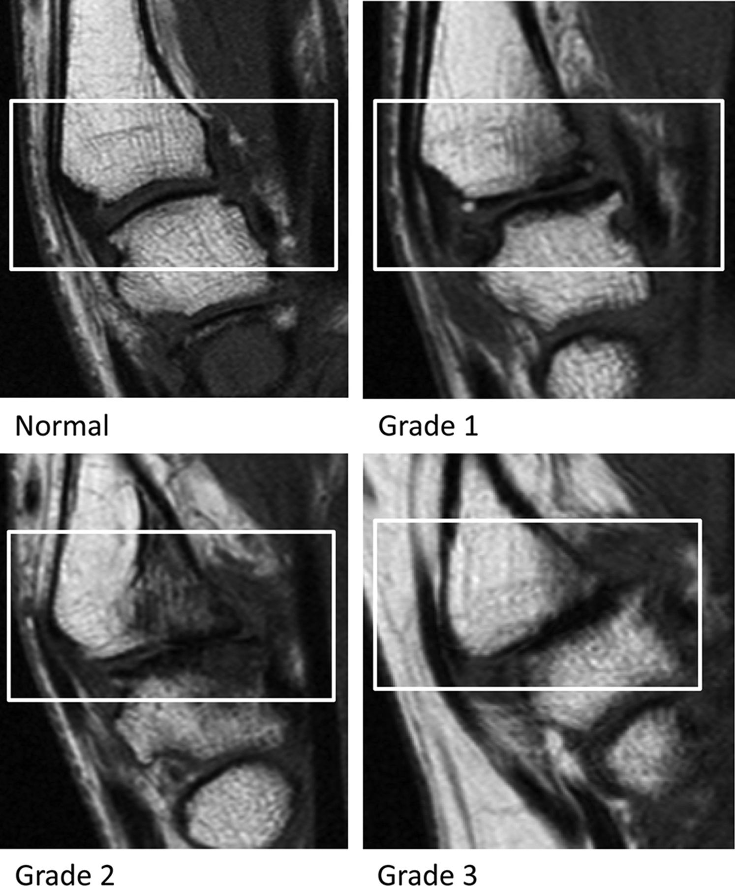
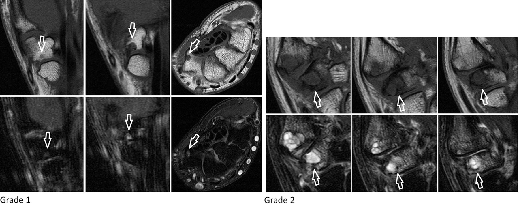
Osteophytes first carpometacarpal: proximal first metacarpal (T1 weighted images).

Osteophytes first carpometacarpal: distal trapezium (T1 weighted images).
Osteophytes scapho-trapezio-trapezoid: proximal trapezium (T1 weighted images).

Osteophytes scapho-trapezio-trapezoid: proximal trapezoid (T1 weighted images).

Osteophytes scapho-trapezio-trapezoid: distal scaphoid (T1 weighted images).
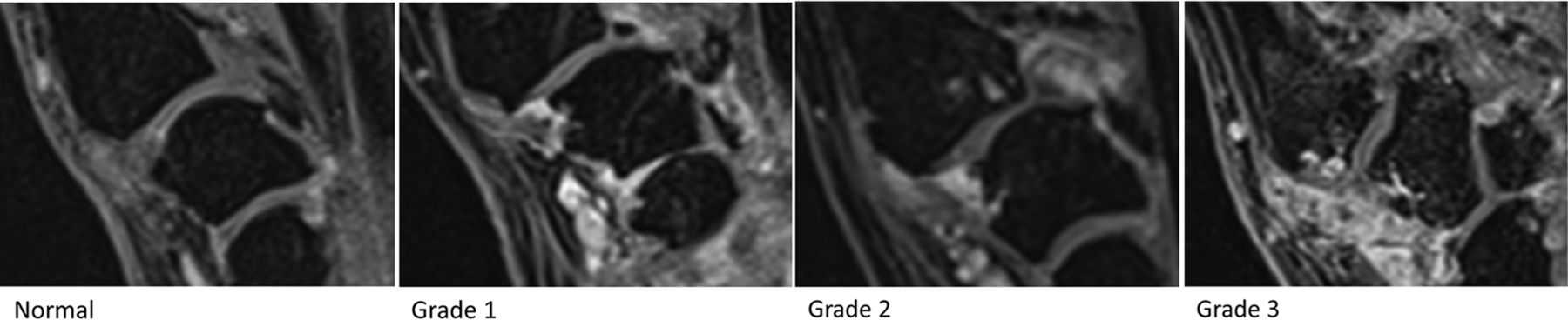
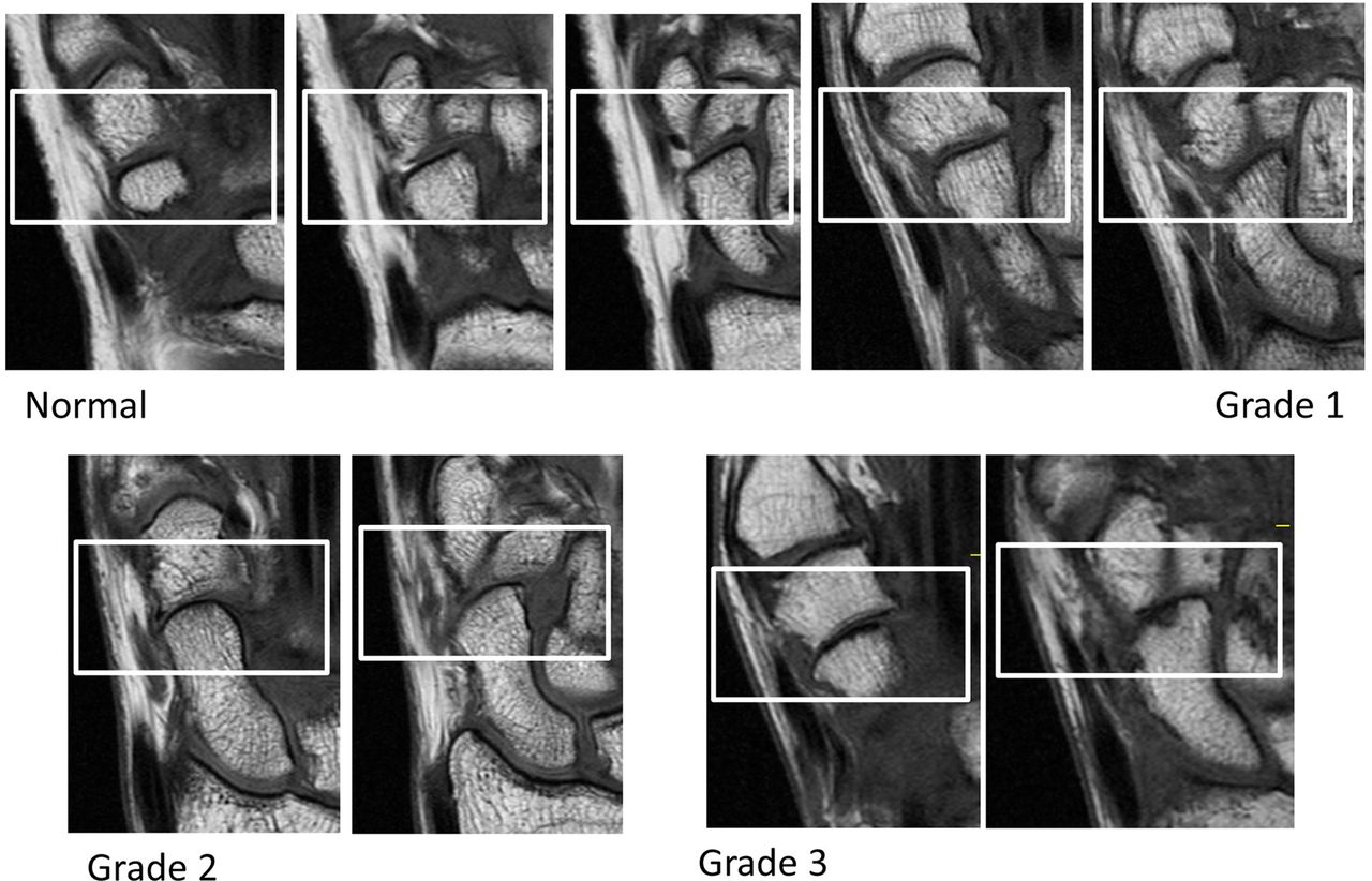
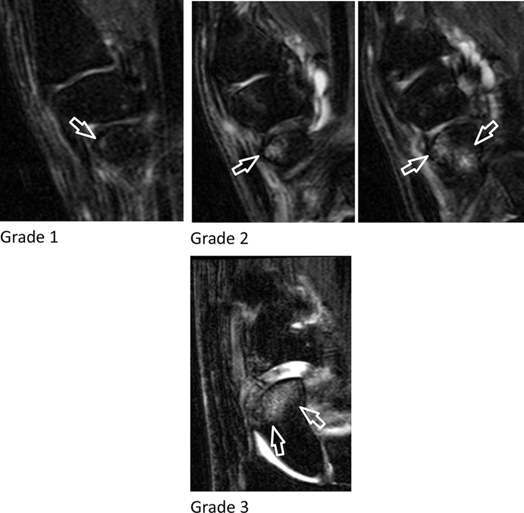
Cartilage assessment first carpometacarpal (T1 weighted images).
Cartilage assessment scapho-trapezio-trapezoid (T1 weighted images).
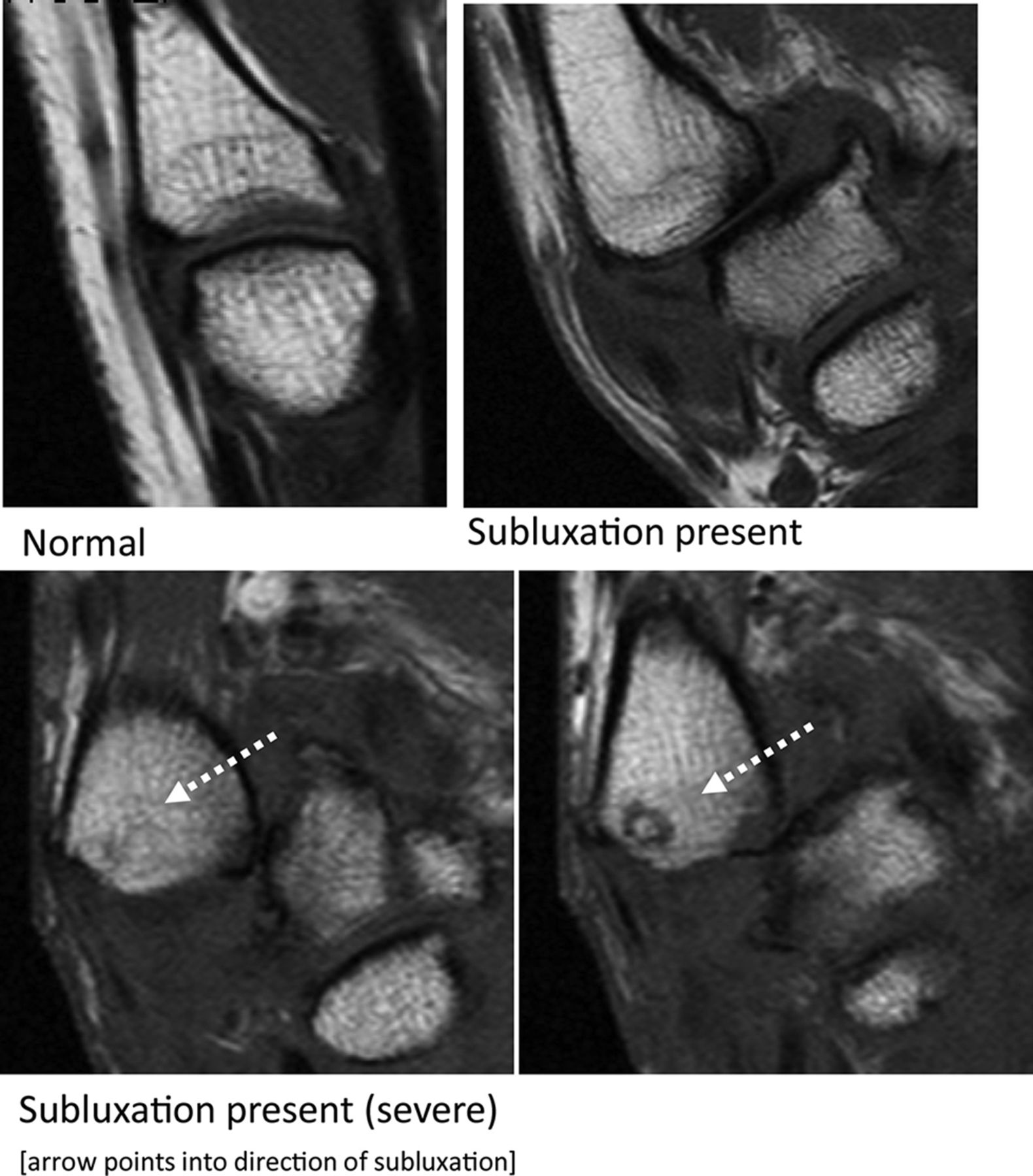
Subluxation first carpometacarpal (T1 weighted images).

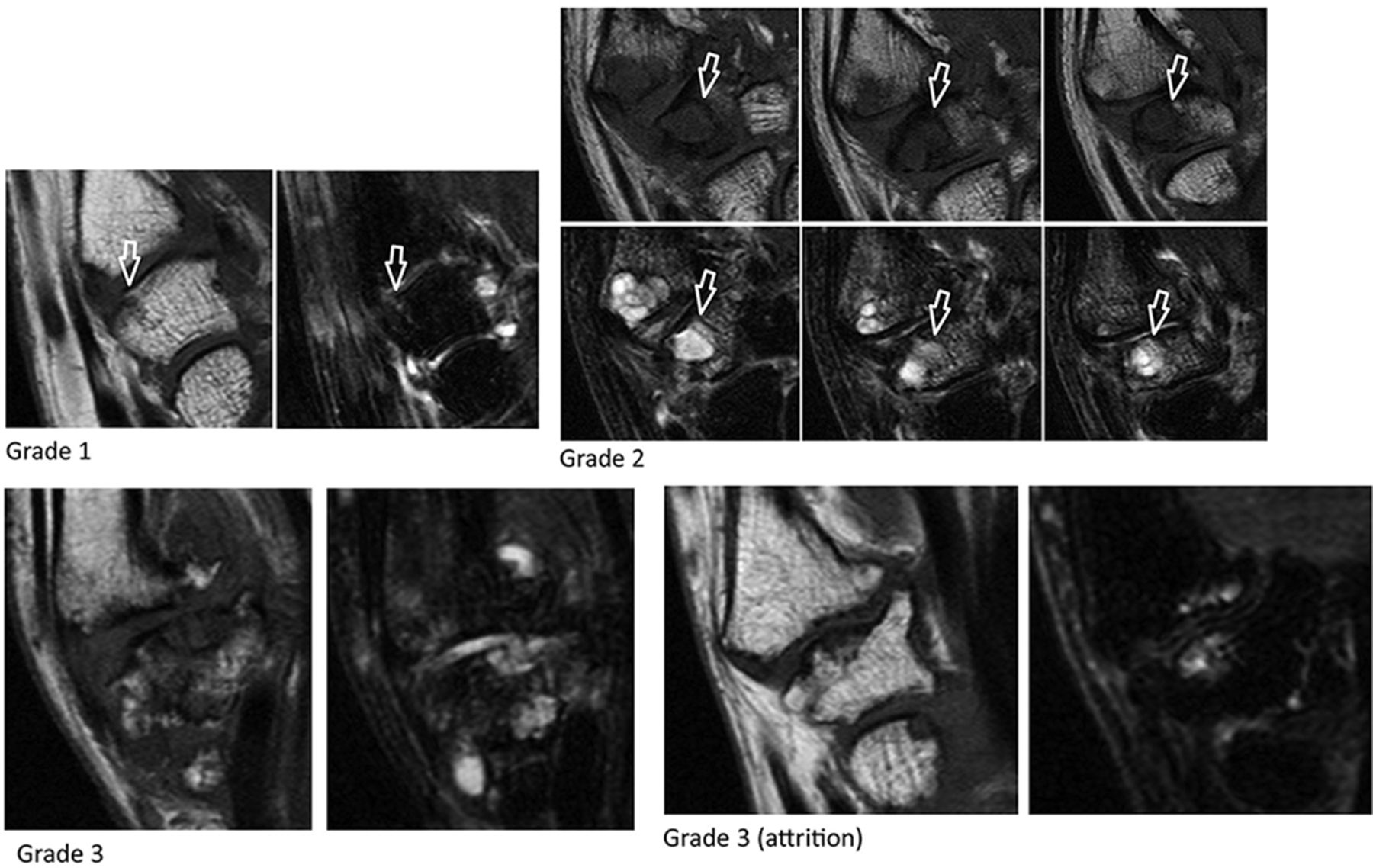
Bone marrow lesions first carpometacarpal: proximal first metacarpal (T2 weighted-fat saturated images).

Bone marrow lesions first carpometacarpal: distal trapezium (T2 weighted-fat saturated images).
Bone marrow lesions scapho-trapezio-trapezoid: proximal trapezium (T2 weighted-fat saturated images).
Bone marrow lesions scapho-trapezio-trapezoid: proximal trapezoid (T2 weighted-fat saturated images).

Bone marrow lesions scapho-trapezio-trapezoid: distal scaphoid (T2 weighted-fat saturated images).
Acknowledgments
Images from the HOSTAS study were provided by the Department of Radiology and Rheumatology of the LUMC (Leiden, the Netherlands). We acknowledge W Damman and R Liu for acquisition of the images. Images from the Nor-Hand study were provided by the Diakonhjemmet Hospital (Oslo, Norway).
Reference
- 1.↵
Footnotes
Contributors FPBK and MK were responsible for the conception of the study. FPBK and IKH were responsible for data acquisition. FPBK, CGP, MR, JLB, IKH and MK were responsible for data analysis, interpretation and drafting of the manuscript. All authors critically revised the manuscript and approved the final version.
Competing interests None declared.
Provenance and peer review Not commissioned; externally peer reviewed.
Data sharing statement FPBK and MK have access to all data, and these are available upon request.
Correction notice This article has been corrected since it first published. The article type has been changed from ’Review' to ’Original article'.