Article Text

Abstract
Background The aim of this work was to summarise the literature evaluating the impact of biopsy procedures, tissue handling, tissue quality and disease-specific aspects including joint biopsied and disease stage, on synovial tissue outcome.
Methods Two reviewers independently identified eligible studies according to the Patients, Intervention, Comparator and Outcome framework obtained for five research questions formulated during the first EULAR task force meeting to produce points to consider (PtC) for minimal reporting requirements in synovial tissue studies. The databases explored were Medline, Embase, CENTRAL and Cinhal. The risk of bias of each study was evaluated using an adapted version of the Joanna Briggs Institute checklist for analytical cross-sectional studies.
Results Of the 7654 records yielded, 75 full texts were assessed, leading to the inclusion of 26 manuscripts in the systematic literature review (SLR). Two papers assessed the impact of biopsy procedures on the quality and quantity of tissue retrieved alongside patient tolerability; six papers focused on synovial tissue variability. Four papers studied the impact of sample handling or randomisation and 14 assessed the impact of disease stage and state, namely early or established active rheumatoid arthritis and remission on histopathological and transcriptomic results.
Conclusions This SLR informs the EULAR PtC for minimal reporting requirements in synovial tissue research in rheumatology. Characteristics related to the study design, population, sample handling, randomisation and analysis can affect the final synovial tissue outcome in the studies reviewed. Thus, accurate reporting of these factors is required in order to ensure the scientific validity of manuscripts describing synovial tissue outcomes.
- arthritis
- rheumatoid
- inflammation
- synovitis
Data availability statement
All data relevant to the study are included in the article or uploaded as supplementary information. Not applicable.
This is an open access article distributed in accordance with the Creative Commons Attribution Non Commercial (CC BY-NC 4.0) license, which permits others to distribute, remix, adapt, build upon this work non-commercially, and license their derivative works on different terms, provided the original work is properly cited, appropriate credit is given, any changes made indicated, and the use is non-commercial. See: http://creativecommons.org/licenses/by-nc/4.0/.
Statistics from Altmetric.com
Key messages
What is already known about this subject?
Synovial tissue biopsy involves different procedures: ultrasound-guided portal and forceps, ultrasound-guided needle biopsy and arthroscopic biopsies.
While different sampling, handling and analysis techniques are reported in publications, their impact on study outcomes remain unknown.
What does this study add?
Histological, immunopathological and transcriptomic outcomes of synovial tissue vary across disease stage and activity.
Different synovial biopsy techniques, provided they involve guidance, do not substantially affect tissue quantity or quality and patient tolerance.
Intra-articular and interarticular variability appears minimal for histopathological or transcriptomic outcomes in rheumatoid arthritis, if number of sample fragments taken from each joint is adequate.
How might this impact on clinical practice?
Information related to tissue retrieval, handling, quality and analysis can affect synovial tissue outcome and therefore should be mandatorily reported in scientific manuscripts.
Introduction
Studies involving synovial tissue (ST) in rheumatic and musculoskeletal diseases (RMDs) have underpinned major breakthroughs in the field of immunology over the past years.1–5 This, in addition to the development of mini-invasive biopsy techniques, has led to a significant increase in synovial biopsy (SB) procedures across centres in Europe and beyond.6–11 While SB procedures are becoming widespread, high standards in both retrieving and analysing ST are required in order to allow a robust evolution of the field. An unmet need exists for evidence and consensus-based points to consider (PtC) defining minimum reporting requirements that could ensure interpretability of the research. In this context, the european alliance of associations for rheumatology (EULAR) has approved the constitution of a task force to develop PtC for minimal reporting requirements in ST research in RMDs.
Although it is assumed that several aspects related to ST handling in research can impact the scientific outcomes, the published studies assessing the influence of tissue retrieving, handling and analysis methods on the results remain scarce and sometimes contradictory. In this context, it was felt important to summarise the existing literature describing how aspects of study design and methods can impact the results. In addition, it is unclear how the joint selected for biopsy, the area within the joint where the tissue is retrieved or how samples are randomised to each analysis, potentially affect the outcome of the research. Therefore, the aim of this systematic literature review (SLR) was to identify the impact of biopsy procedures, tissue handing, tissue quality and disease-specific aspects, on tissue outcomes, to inform the EULAR PtC for minimal reporting requirements in ST research in RMDs.
Methods
Research questions
The SLR was conducted following the EULAR standardised operating procedures.12 The scope of the literature search was defined during a first virtual task force meeting, in which five different research questions (RQ) were formulated and approved by all task force members and focused on biopsy techniques (RQ1), representativity of ST from large and small joints in inflammatory RMDs (RQ2), sample randomisation and tissue handling (RQ3), tissue quality control (RQ4) and impact of disease stage and activity on tissue outcome (RQ5).
Search methodology
The five RQs were transformed using the ‘Patients, Intervention, Comparator and Outcome’ (PICO) framework to determine the search strategy (online supplemental text S1).13 The search was run by an experienced librarian from Paris University, Paris, France (CW). The keywords selected and used for each PICO and databases are presented as supplementary material (online supplemental text S2). The databases explored were Medline, Embase, CENTRAL and Cinhal. Hand search for individual original research studies and crosscheck for references from specific rheumatology and immunology journals were performed.
Supplemental material
Study selection, data extraction and risk of bias assessment
Titles and abstracts of the retrieved papers were assessed by two independent reviewers (AN and FC). General eligibility criteria were described as follows: original research articles, published in peer-reviewed journals, English language, reporting clinical or translational research involving ST. Cohen’s kappa agreement between reviewers was 0.95. Discrepancies were resolved by discussion.
Data on patients’ characteristics, scientific methods of analysis, parameters assessed and outcomes were extracted. In addition, all manuscripts included in the final SLR were assessed from their content against criteria compelled during the first task force meeting by the task force members. These criteria identified important areas where data should be reported on a mandatory basis.
Due to the lack of validated risk of bias (RoB) tool for translational research in rheumatology, the RoB was assessed using an adapted version of the Joanna Briggs Institute checklist for analytical cross-sectional studies.14 Briefly, eight questions related to specific methodological aspects were applied to the study. If ≥2 ‘no’ answers were returned, the study was considered at high RoB, while RoB was said to be ‘unclear’ if one ‘no’ answer was returned. The tool is detailed in online supplemental text S3.
Results
Study selection and study characteristics
Overall, our search yielded 7654 abstracts out of which 2181 duplicates were excluded; 5473 titles and abstracts were screened, and 75 full texts assessed, leading to the inclusion of 26 manuscripts in the SLR. Additional details are presented in the flow chart (figure 1). Study characteristics with their type, objective, number and characteristics of patients and studied outcomes are presented in table 1. The RoB assessment is presented in table 2. Most studies were found to have an unclear (n=14) or high (n=12) RoB mainly due to the existence of confounding factors.
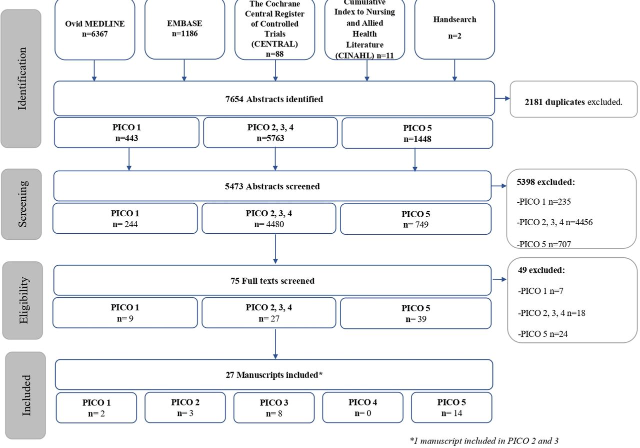
Flow chart summary of the systematic literature review, article identification, screening and final selection. PICO, patient, intervention, comparison, outcome.
Design, outcomes and population of studies included in the manuscript
Risk of bias assessment of all included studies
Reporting methods and outcomes across included studies
We additionally analysed the way each included manuscript reported their materials and methods section, with a focus on several areas deemed important by the task force, as listed in table 3. Essentially, manuscripts have been checked for the following criteria: clinical data, biopsy procedure, tissue handling, tissue quality control and tissue outcomes. The area most likely to be reported in all manuscripts were: demographics (100%), disease duration (92%), target joints for biopsy (81%), number of fragments retrieved and included for analysis (73%) and their processing (100%), scoring/quantification system used for immunohistochemistry (IHC, 96%) and disaggregation method used for single-cell analysis (100%). The least likely items to be reported (0%–40%) were: ultrasound (US) features of the biopsied joints (for US-guided procedures), operator’s experience and adverse events related to the biopsy procedure, intraobserver or interobserver reproducibility for IHC and RNA quantity, purity and quality control method.
Reporting of different areas pertaining to synovial tissue sampling, handling or analysing
Impact of the biopsy procedure and device on tissue quality and biopsy tolerability
One study with unclear RoB assessed the impact of biopsy procedure and devices on ST yield and quality. In this study, the three main guided biopsy techniques: US-guided needle biopsy (NB), ultrasoundUS-guided portal and forceps (P&F), arthroscopy were compared with each other and with blind NB. The number of graded ST fragments/total number of ST fragments was higher in guided techniques compared with blind NB for both small and large joints although this result was only significant for large joints (p=0.048 and p=0.057, respectively).15 While all guided techniques enabled retrieval of a sufficient amount and quality of tissue for a meaningful analysis, large joint arthroscopic-guided biopsy (A) yielded higher quantities of RNA than US-guided NB and US P&F biopsy (A vs NB p=0.002, A vs P&F p=0.0014) and higher RIN for A compared with NB (p=0.0018) but not compared with P&F (p=0.068). No difference was observed in immune cell infiltrate and pathotype regardless of the technique used. For sequential biopsies performed with any of the guided techniques, no difference was observed between first and second biopsies for tissue yield and quality or RNA quality.
As far as tolerance is concerned, one study with unclear RoB assessed the influence of the biopsy technique on patient-reported outcomes.16 Visual analogic scale (VAS) for pain, swelling and stiffness did not differ for A, NB or P&F, while the willingness to repeat was superior in P&F compared with both A (p<0.01) and NB (p<0.01).16 In addition, no difference was observed in terms of reported PROs for tolerance or adverse events between first and second biopsies although numbers were low.
These results are summarised in tables 4 and 5 (sequential biopsies).
Tissue outcome and tolerance of synovial biopsies
Tissue outcome and tolerance of sequential synovial biopsies
Impact of the joint biopsied on tissue outcomes
Two studies with high RoB17 18 and one with unclear RoB19 assessed the impact of the joint biopsied on either cell infiltrate,17 19 transcriptomics19 or T-cell repertoire (TCR), deemed to be particularly relevant in ST arthritis of autoimmune mechanisms.18 19 Of interest, when comparing outcomes from tissues retrieved in small versus large joints from the same patients with rheumatoid arthritis (RA), the authors could not identify any difference in immune cell infiltrate and IL-6 expression assessed by non-parametric paired analysis.17 In addition, correlations were found for CD68 +macrophages, CD3 +T cells, CD138 +plasma cells and IL-6 expression and CD68 +macrophages and CD3 +T cells in the ST sublining of both small and large joints.17 As far as the synovial lining layer is concerned, significant correlations were found between small and large joints for lining layer hyperplasia, inflammatory infiltrate in H&E staining.19 The same transcripts were shown to be overexpressed in both small and large joints, and the same T-cell clone repartition was observed in small and large and right and left joints of the same individuals with RA.18 19
It is noteworthy that all studies included small numbers of patients and, therefore, the absence of difference could also reflect a lack of power rather than a true similarity.
Impact of tissue handling and sample randomisation on tissue outcomes
One article with unclear RoB20 assessed the impact of ST handling on quality and outcomes. They compared different tissue preservation (fresh vs cryopreserved tissue) and dissociation methods (mechanical vs mechanical +enzymatic) in tissue obtained from n=16 US-guided NB and P&F performed in RA patients and/or surgical biopsies performed n=32 RA patients. Interestingly, the cryopreservation did not impact the viable cell count or repartition of synovial cell subpopulations assessed by flow cytometry compared with freshly processed tissue. On the other hand, the mechanical +enzymatic technique using Liberase retrieved a higher total cell count per gram of ST, higher viable cell count and better repartition of cell subpopulations.
Few studies specifically assessed intra-articular variability. Authors retrieved ST from different compartments of the knee joint using arthroscopy, including comparisons of cartilage pannus-junction (CPJ) and supra-patellar recess (SP),21 lateral, medial and SP recesses versus CPJ22 and SP versus infra-patellar (IP) recess.18 The immune cell infiltrate assessed by IHC showed no major difference among the different compartments of the knee, including CD68+, CD3+, CD4+, CD8+, CD22+, CD38 + cells21 and CD45RO+, CD3+, CD8 + and Mac 387+ cells.22 These findings need to be interpreted carefully due to a low number of samples leading to a possible lack of power. Granzyme B and matrix metallo-proteinases (MMP-1, MMP-3 and MMP-13) expression was also assessed by IHC showing no differences.21 In addition, IL-1β, TNF, IL-2, IL-4, IL-6, IL-8 and IFN γ levels were correlated in all knee recesses, while IL-10, granulocyte-macrophage colony stimulating factor (GM-CSF) did not appear correlated across knee compartments.22 The TCR repertoire study showed no difference in the most frequently expressed T-lymphocyte clones across SP and IP compartments.18
Finally, a few studies with high23 24 and unclear RoB15 25 studied sampling error by evaluating the minimum number of tissue samples needing to be included in a single analysis to obtain consistent results. Of note, two of these studies assessed ST retrieved from arthroplasty surgery and not synovial biopsies. In small joints, it was demonstrated that the semiquantitative IHC analysis of four tissue fragments provided a reliable sample analysis with 10% sampling error (mean difference <10% in 91.6% of the cases).11 If quantitative analysis is used, the analysis of a cumulative area of 2.5 mm2 and/or six (large joints) to eight (small joints) randomly selected sections (from different samples or from different cutting levels) allowed a mean difference <10% in 100% of cases.23 25 As far as cytokine expression (IL-1β, IL-6, TNF-α and MMP-1) by PCR in large joints is concerned, four to seven tissue fragments were deemed necessary in order to detect a twofold change with a 25% sampling error.24
Impact of the disease stage on the tissue outcomes
Fourteen studies assessed the impact of disease stage on histopathological findings in RA,26–33 osteoarthritis (OA)34 or transcriptomics aspects in RA,35–38 including eight studies with high RoB and seven with unclear RoB.2 27 31–34 37 Of interest, while contradictory results for lining layer thickness and CD68 +cell infiltrate (lining layer and sublining) in early versus established RA were reported, other cell infiltrates (CD3, CD4, CD8, CD20 or CD22, CD38 and mast cells) and vascularity in sublining appeared to be the same between early and established RA.26 39 In addition, proinflammatory cytokines (TNFα, IL- 1Beta, IL-6 and MCP-1), MMPs, toll-like receptors 2, 3 and 4 and integrins alphaVBeta5 and 3 did not appear to be expressed differentially in STs across disease stages,26 29 39 40 while cells displaying apoptotic features and p53 positive cells were increased in established RA.28 30 Another study has compared histopathological infiltrate between knee synovitis in patient with active naïve RA and US power Doppler negative knee synovitis in patients with RA in remission or low disease activity. Interestingly, authors showed a significantly higher infiltrate of CD68+, CD20+, CD21 + and CD3+ cell in ST from naïve RA patients.31 Two other studies interrogated ST infiltrate in early naïve versus resistant established RA versus RA in remission.32 33 In one study, the authors report that the Krenn Synovitis Score (KSS) was higher in patients with early active than established RA and both were higher compared with patients in remission.32 In addition, established RA resistant to TNFi had higher KSS and higher prevalence of B cell–rich synovitis than early untreated RA.33 It is important to note that the definition of ‘early’ and ‘established’ RA was variable across studies and this might explain the contradictory results observed. Detailed results are summarised in tables 6 and 7.
Impact of disease stage on histopathological tissue outcomes in RA
Impact of disease stage on histopathological tissue outcomes in OA
As far as OA is concerned, Ostojic et al have described an increased lining layer thickness and stromal infiltration in mild OA (Kellgren and Lawrence Score (KLS) 1–2) compared with advanced OA (KLS 3–4), while cell infiltrate was more pronounced in advanced OA compared with mild. Overall, the KSS was superior in mild OA suggesting synovitis in early stages compared with more advanced and severe OA (table 7).34
Finally, four studies looked into transcriptomic expression across the RA spectrum. The two studies from Guo et al showed no difference in expression of genes related to CD40–CD40L36 or PD-1 PD-L135 pathways activity in STs from 57 early RA (<1 year) versus 95 established RA (>1 year). Lequerré et al showed differing transcriptomic signatures in a lower number of samples (n=3 early RA (<9 months) and n=4 established RA (>4 years)).37 Early RA samples overexpressed 503 genes corresponding to 12 biological processes, including immunity and host defenses, stress responses, T-cell-mediated immunity and tumour suppressor and MHC class II mediated immunity; while established RA tissues overexpressed 216 genes linked to the following biological processes: cell cycle, cell surface receptor-mediated signal transduction, cell cycle control, ligand-mediated signalling, apoptosis inhibition and granulocyte-mediated immunity. In addition, Alivernini et al assessed distinct ST macrophage clusters using single-cell RNAsequencing in tissues collected by US-guided biopsies across different RA phases (naive RA vs established RA vs remission RA, respectively). Of interest, MerTK +clusters (TREM2high and LYVE1pos) and increased proportions of MerTK-CD48-SPP1+ and MerTK-CD48-S100A12+ clusters were observed in naïve and resistant RA, while ST from patients with RA in remission were characterised by an increase in the MerTKposFOLR2highLYVE1pos cluster. Overall, these data are suggestive of different ST macrophages clusters with very diverse phenotypes and function acting at different periods of the disease course.38
Discussion
This SLR summarises the available evidence on the impact of several aspects of SB methods on tissue outcomes. To the best of our knowledge, this is the first systematic approach examining the impact of a wide range of methods on the results of ST assessment, whether it is histological, bulk transcriptomic or more advanced single cell technologies. We were able to retrieve a number of studies suggesting that the biopsy technique, regardless of the guiding technique (arthroscopic or US guided) was well tolerated and allowed to retrieve a sufficient amount of tissue for a meaningful analysis, suggesting that any of these techniques can be used for translational research. On the other hand, tissue yield and quality were consistently shown to be reduced with blind NB technique. The analysis also showed that most histopathological and transcriptomic outcomes were similar regardless of the joint biopsied (small vs large, right vs left) or the recess of the joint that was biopsied in the same patient (ie, knee recesses). Of note, all the studies pertaining to this RQ were performed in ST retrieved in RA patients and these results are not necessarily applicable to other RMDs. Similar studies using the most advanced technologies are however warranted, since joint-dependent epigenetic differences among ST resident cells have been reported.41
It is noteworthy that a low number of studies was retrieved to address the different RQs, and one RQ pertaining to the impact of tissue quality on outcomes was not addressed due to the absence of relevant articles. Moreover, it is important to mention that the majority of the studies was performed in patients with RA and in research settings; and those findings may not be necessarily generalisable to other inflammatory conditions since the pathogenetic mechanisms may be different, or other clinical settings. In addition, all the articles had a high or unclear RoB, frequently due to the existence of confounding factors. These biases related mostly to the study design that may affect the results. Therefore, efforts should be undertaken to reduce them. This could be achieved by carefully designing studies with more homogeneous inclusion criteria and better-defined baseline populations. While in the past the paucity of centres with SB expertise and the invasiveness of the procedures were a limiting factor, the implementation of US-guided SB procedures in a larger number of centres, thanks to an improved tolerance and reduced invasiveness, will help in evaluating the real impact of this procedure. In addition, it will allow the development of prospective studies with a higher number of patients better powered to take into account both clinical and tissue heterogeneity.
Although this SLR aimed to assess evidence in both clinical and research settings, the studies referring to clinical practice were extremely scarce. Therefore, the analysis of ST should be reinforced as an area of high interest and studies aimed at defining disease endotypes and their applications, both for research purposes, and also for practicing clinicians, should be prioritised over the coming years. Such studies will contribute to building data sets and evidence supporting the use of ST analysis for standard of care diagnosis, prognosis or disease management.
In conclusion, this SLR retrieved evidence which contributed to inform the EULAR PtC for minimal reporting requirements in ST research in rheumatology.
Data availability statement
All data relevant to the study are included in the article or uploaded as supplementary information. Not applicable.
Ethics statements
Patient consent for publication
Ethics approval
This study does not involve human participants.
References
Supplementary materials
Supplementary Data
This web only file has been produced by the BMJ Publishing Group from an electronic file supplied by the author(s) and has not been edited for content.
Footnotes
Twitter @AurelieRheumo
Contributors All authors contributed to and finally approved the current manuscript. MAd’A and AF share last authorship.AN acts as guarantor.
Funding This work was funded by EULAR (SCI020).
Competing interests AN has received consulting and/or speaker’s fees from UCB, CHUGAI, BMS all unrelated to this manuscript. FC has received consulting and/or speaker’s fees from Lilly, Novartis and UCB, all unrelated to this manuscript. MAD'A has received consulting and/or speaker’s fees from Lilly, Novartis, AbbVie, BMS, Pfizer, Galapagos and UCB, all unrelated to this manuscript. AF has received consulting, research grants and/or speaker’s fees from Abbvie, Roche, Jjanssen, UCB, Nascient, Mestag, GSK, all unrelated to this manuscript.
Provenance and peer review Not commissioned; externally peer reviewed.
Supplemental material This content has been supplied by the author(s). It has not been vetted by BMJ Publishing Group Limited (BMJ) and may not have been peer-reviewed. Any opinions or recommendations discussed are solely those of the author(s) and are not endorsed by BMJ. BMJ disclaims all liability and responsibility arising from any reliance placed on the content. Where the content includes any translated material, BMJ does not warrant the accuracy and reliability of the translations (including but not limited to local regulations, clinical guidelines, terminology, drug names and drug dosages), and is not responsible for any error and/or omissions arising from translation and adaptation or otherwise.