Article Text

Abstract
Objectives To evaluate humoral and cellular immune responses and adverse events (AEs) after COVID-19 vaccination in patients with primary Sjögren’s syndrome (pSS) compared to healthy controls (HC), and disease activity following vaccination in patients with pSS.
Methods 67 patients with pSS and 33 HC (ratio 2:1) received COVID-19 vaccinations following the Dutch vaccination programme. Patients with pSS did not use immunomodulatory drugs, except hydroxychloroquine. Anti-spike 1 receptor binding domain IgG serum antibody levels were measured 28 days after complete vaccination. AEs were collected 7 days after vaccination. In a subgroup, salivary anti-SARS-CoV-2 antibodies and T-cell response by interferon-γ enzyme-linked immune absorbent spot was measured.
Results 47 patients with pSS (70%) and 14 HC (42%) received BNT162b2 (Pfizer-BioNtech), 13 (19%) and 5 (15%) received ChAdOx1 nCoV-19 (AstraZeneca), 6 (9%) and 8 (24%) received mRNA-1273 (Moderna), and 1 (1%) and 6 (18%) received Ad.26.COV2.S (Janssen). All participants had positive anti-SARS-CoV-2 antibody levels (>2500 AU/mL) postvaccination. No differences in anti-SARS-CoV-2 antibody levels were observed between patients with pSS and HC, for each vaccine type. Salivary anti-SARS-CoV-2 IgG antibodies also increased, and a T-cell response was observed in patients with pSS and HC. Frequencies of systemic AEs were comparable between patients with pSS and HC (first vaccination: 34/67 (51%) vs 16/33 (48%), p=0.83; second: 41/66 (62%) vs 14/25 (56%), p=0.59). No significant worsening was observed in patient-reported and systemic disease activity, including auto-antibodies.
Conclusions Patients with pSS had similar humoral and cellular immune responses as HC, suggesting COVID-19 vaccination is effective in patients with pSS. AEs were also comparable, and no increase in disease activity was seen in patients with pSS.
- COVID-19
- Vaccination
- Sjogren's Syndrome
Data availability statement
No data are available. Currently there are no plans to share additional data beyond what is included in this article.
This is an open access article distributed in accordance with the Creative Commons Attribution Non Commercial (CC BY-NC 4.0) license, which permits others to distribute, remix, adapt, build upon this work non-commercially, and license their derivative works on different terms, provided the original work is properly cited, appropriate credit is given, any changes made indicated, and the use is non-commercial. See: http://creativecommons.org/licenses/by-nc/4.0/.
Statistics from Altmetric.com
Key messages
What is already known about this subject?
Patients with primary Sjögren’s syndrome (pSS) worry about the effectiveness and possible side effects of COVID-19 vaccination.
Few data is available on the effectiveness and side effects of COVID-19 vaccination in patients with pSS.
What does this study add?
Patients with pSS had similar levels of anti-SARS-CoV-2 antibodies compared with healthy controls (HC), providing evidence that COVID-19 vaccination is effective in patients with pSS.
Adverse events were comparable between patients with pSS and HC and no increase in disease activity was seen in patients with pSS.
How might this impact on clinical practice or further developments?
Our findings are an important and reassuring message to patients with pSS and their treating physicians, and support the use of COVID-19 vaccinations in patients with pSS. Furthermore, our data provide arguments that patients with pSS may also benefit from future booster vaccinations.
Introduction
Patients with primary Sjögren’s syndrome (pSS) worry about the effectiveness and possible side effects of COVID-19 vaccination, in particular flaring of the disease.1 Certain factors may contribute to their concerns, such as an imbalance of the immune system (eg, lymphocytopenia), severe organ manifestations or in some cases use of immunosuppressive drugs.2 Most previous studies investigated COVID-19 vaccination response in patients with various rheumatic diseases who were on immunosuppressive drugs.3–5 However, only few patients with pSS were included in these studies and little is known about COVID-19 vaccination responses in patients with pSS without immunosuppressive drugs, which is the case for the majority of this patient population. pSS is a systemic, auto-immune disease, which is generally characterised by an over-active immune system, illustrated by B-cell hyperactivity.6 Previous studies showed that untreated patients with pSS produced higher influenza-specific antibody levels after influenza vaccination compared with healthy controls (HC).7 8 Influenza vaccinations also resulted in concomitant elevation of anti-EBV and auto-antibody levels, an indication of polyclonal B-cell activation.7 8 Additionally, influenza-specific antibody levels were associated with higher steady-state interferon (IFN) signatures in monocytes.7 Potentially, the toll-like receptor-7 (TLR-7)/type-I IFN pathway is an important driver of polyclonal B-cell activation in pSS.9 A type-I IFN signature is present in approximately 55%–80% of patients with pSS, and has been associated with anti-SSA positivity and higher haematological and biological activity.9–11 Furthermore, TLR-7 is overexpressed in patients with pSS.12 For SARS-CoV-2 host defence, this pathway seems particularly important, as illustrated by a study showing that four young males with live-threatening COVID-19 infection, without pre-existing medical conditions, had a loss-of-function TLR-7 variant on their X-chromosome and functional defects in type-I and type-II IFN responses.13
Based on these findings, we hypothesised that patients with pSS may develop a stronger humoral response to COVID-19 vaccination. On the other hand, possible vaccination-induced polyclonal B-cell activation might lead to more side effects, or patients may experience an increase in disease activity after vaccination. Therefore, the primary objectives of this study were to evaluate anti-SARS-CoV-2 antibody responses in serum and adverse events (AEs) after COVID-19 vaccination in patients with pSS compared with HC. A secondary objective was to evaluate disease activity after vaccination in patients with pSS. Cellular response, anti-SARS-CoV-2 antibody levels in saliva and auto-antibody levels in serum were also measured.
Methods
Study design
This study is a prospective, single-centre, longitudinal cohort study conducted in the pSS expertise centre at the University Medical Centre Groningen (UMCG; Groningen, Netherlands). Patients with pSS and female HC were included in a 2:1 ratio. Inclusion criteria were age of 18–75 years and exclusion criteria were a PCR-confirmed SARS-CoV-2 infection (current/previous) and pregnancy. Patients with pSS had to fulfil the ACR/EULAR classification criteria for pSS14 and additional exclusion criteria were current use of conventional or biological DMARDs (except hydroxychloroquine) or prednisone >10 mg/day, and previous use of DMARDs ≤6 months before inclusion (rituximab ≤12 months). For HC, additional exclusion criteria were presence of auto-immune or mixed connective tissue diseases, confirmed infectious, inflammatory or malignant disease, and use of immunosuppressive medication.
Participants received COVID-19 vaccinations following the Dutch vaccination programme. Vaccine types included in the vaccination programme were BNT162b2 (Pfizer-BioNtech), ChAdOx1 nCoV-19 (AstraZeneca), mRNA-1273 (Moderna) or Ad.26.COV2.S (Janssen). Participants received two doses of the Pfizer-BioNtech, AstraZeneca or Moderna vaccine, or one dose of the Janssen vaccine. At the start of this study (March 2021), the time interval between the doses were 6 weeks for Pfizer-BioNtech, 12 weeks for AstraZeneca and 4 weeks for Moderna, but this varied during this study.
Study measurements
Baseline demographics were collected from electronic patient files for patients with pSS and from questionnaires for HC. Blood was collected by a finger prick at home. Spike 1 (S1)-receptor binding domain (RBD)-neutralising IgG antibodies were measured using Siemens Healthineers antibody assay (Labonovum, Limmen, the Netherlands).15 A level of >2500 AU/mL was considered positive. Total serum levels of IgG were also measured. The finger prick was collected before the first vaccination, to confirm that participants did not have a previous SARS-CoV-2 infection, and 28 days after the final vaccination.
Incidence and severity of (serious) AEs were self-reported 7 days after each vaccination. Frequencies of AEs were also compared with a general population cohort of the Dutch pharmacovigilance centre Lareb.16 For patient-reported disease activity, EULAR Sjögren’s Syndrome Patient-Reported Index (ESSPRI) and patient global disease activity were collected at baseline and 28 days after complete vaccination. Systemic disease activity measured with EULAR Sjögren’s Syndrome Disease Activity Index (ESSDAI) was retrieved from medical records ≤1 year before the first vaccination (median time 4 months) and ≤6 months after the first vaccination.
Subgroup measurements
All participants (pSS and HC) were invited to donate additional blood and/or saliva samples. This was not mandatory for participation in this study. Additional blood samples were drawn by venipuncture before vaccination and 7 days after the second vaccination (14 days after the first for Janssen) and stimulated whole saliva (paraffin-chewing; 5 min) was collected before vaccination and 28 days after complete vaccination.
To validate the finger prick antibody levels, anti-S1 and anti-RBD IgG levels were measured in postvaccination blood samples using the multiplex immuno assay (MIA) at the National Institute for Public Health and the Environment (RIVM).17
In the prevaccination and postvaccination blood samples, frequencies of SARS-CoV-2 spike-specific T-cells were assessed by IFN-γ enzyme-linked immune absorbent spot (ELIspot) assay.18 To calculate spike-specific T-cell response, the average spot-forming cell (SFC) count of the negative control was subtracted from summed average of the S1 and S2 SFC counts. Change (Δ) from prevaccination to postvaccination spike-specific SFC counts was calculated. Responders were defined as patients who had at least a twofold increase in postvaccination compared with prevaccination SFC counts and SFC counts of ≥50/106 cells in the postvaccination sample.
In patients with pSS, anti-SSA antibody levels (Ro52 and Ro60) were measured in prevaccination and postvaccination blood samples by an in-house ELISA.19
In the saliva samples, anti-S1 and anti-RBD IgG and IgA antibody levels were measured using a fluorescent-bead-based MIA.20
Statistical analyses
Descriptive statistics were used for baseline characteristics and safety outcomes. Results were expressed as number (%), mean±SD or median (IQR) for respectively categorical, normally or non-normally distributed data. Differences between groups were tested using χ2 test or Fisher’s exact test for categorical data, independent samples t-test for normally distributed data and Mann-Whitney U test for non-normally distributed data. For changes within patients over time paired t-test was used for normally distributed data and Wilcoxon signed-rank test for non-normally distributed data. For correlations, Spearman’s correlation test was used. Univariate linear regression was performed with SARS-CoV-2 antibody level as dependent variable and pSS/HC group as independent variable. Multivariate linear regression was used to correct for age as potential confounder. SARS-CoV-2 antibody levels were log-transformed to obtain a normal distribution of residuals in the linear regression models. Within the pSS group, univariate linear regression was used to explore associations between SARS-CoV-2 antibody levels and baseline characteristics. Main analyses were split for the separate vaccine types. P values <0.05 were considered statistically significant. Statistical analyses were performed using SPSS V.23.0.
Results
Baseline characteristics
One patient was excluded because of a positive prevaccination anti-SARS-CoV-2 level of 59113 AU/mL and symptoms fitting with COVID-19 in March 2020. Two patients with pSS had a (low) positive anti-SARS-CoV-2 antibody level before vaccination (3470 and 2628 AU/mL). Both patients did not report a previous COVID-19 infection or positive PCR test for COVID-19, had negative anti-Nucleocapsid (N) antibodies and no spike-specific T-cell response prevaccination. Therefore these patients were included.
In total, 67 patients with pSS and 33 HC were included. Following the Dutch national vaccination programme, the majority of patients and HC received the Pfizer-BioNtech vaccine (pSS: 47 (70%), HC: 14 (42%)), or AstraZeneca (pSS: 13 (19%), HC: 5 (15%)). Six (9%) patients with pSS and 8 (24%) HC received Moderna, and 1 (1%) pSS patient and 6 (18%) HC received Janssen. Overall, patients with pSS were significantly older compared with HC, which was mainly attributed to the younger age in HC who received Moderna or Janssen vaccines (table 1). In the Pfizer-BioNtech and AstraZeneca groups, no differences in age between patients with pSS and HC were observed (table 1). At baseline, 58 (87%) patients with pSS were female, 15 (22%) used hydroxychloroquine, no patients used prednisone, and median ESSDAI was 3 (IQR 1–4).
Baseline characteristics of patients with pSS and healthy controls
In 31 patients with pSS and 11 HC additional blood and/or saliva samples were collected. Baseline characteristics of this subgroup were comparable to those of the total group (online supplemental table 1).
Supplemental material
SARS-CoV-2 antibody levels in serum
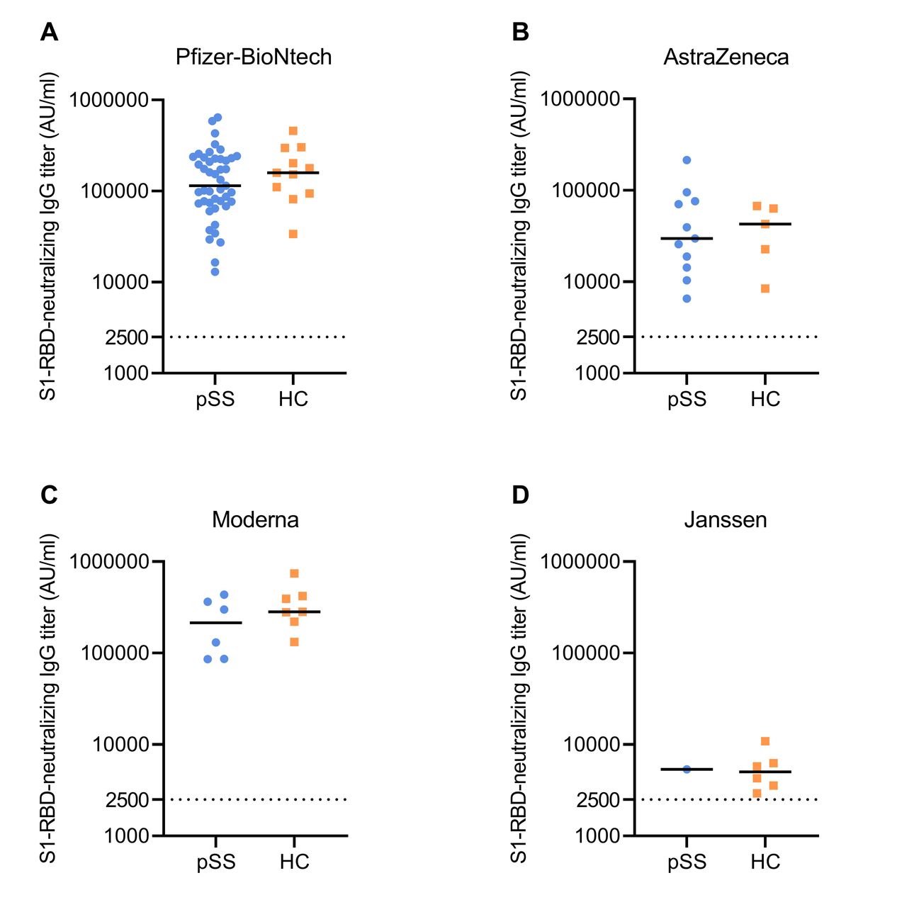
Postvaccination finger prick antibody levels were available from 63 patients with pSS and 29 HC. The finger prick and RIVM antibody levels showed a good correlation: r=0.787 (p<0.001) for anti-S1 and r=0.785 (p<0.001) for anti-RBD IgG (n=36). All participants had positive anti-SARS-CoV-2 IgG levels postvaccination. Because large variation was seen in antibody levels between the various vaccine types, analyses were split per vaccine type. No significant differences in anti-SARS-CoV-2 antibody levels were observed between patients with pSS and HC for all vaccine types (figure 1). Using multivariate linear regression, we found no confounding effect of age on SARS-CoV-2 antibody levels in the pSS versus HC group for the Pfizer-BioNtech or Moderna vaccines. For the AstraZeneca group, age was identified as confounder, but correcting did not lead to significant differences in antibody levels (online supplemental table 2). The Janssen group was underpowered for this analysis. Within the pSS group, univariate linear regression did not show a significant effect of baseline hydroxychloroquine use, total serum IgG, lymphocyte count or ESSDAI on anti-SARS-CoV-2 antibody levels both for participants who received Pfizer-BioNtech and AstraZeneca (online supplemental table 3). The Moderna and Janssen group were too small for such analyses.

Finger prick SARS-CoV-2 antibody levels (S1-RBD neutralising IgG) of participants who received (A) Pfizer-BioNtech (B) AstraZeneca (C) Moderna or (D) Janssen. An antibody level of ≥2500 AU/mL was considered positive, indicated with the dashed line. HC, healthy controls; pSS, primary Sjögren’s syndrome; RBD, receptor binding domain; S1, spike 1.

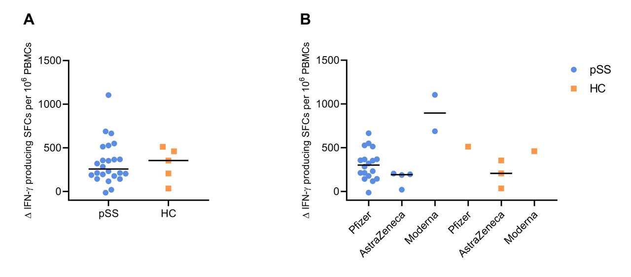
T-cell response measured with IFN-γ ELIspot assay for (A) total group and (B) split per vaccine type. The change (Δ) in IFN-γ producing SFCs from prevaccination to postvaccination is presented. ELIspot, enzyme-linked immune absorbent spot; HC, healthy controls; IFN, interferon; PBMC, peripheral blood mononuclear cell; pSS, primary Sjögren’s syndrome; SFC, spot-forming cell.
T-cell response
The IFN-γ ELIspot assay for T-cell response was only performed in patients who received Pfizer-BioNtech, AstraZeneca or Moderna vaccines (pSS: n=24, HC: n=5) and from whom peripheral blood mononuclear cells were collected. 20/24 (83%) patients with pSS and 4/5 (80%) HC were responder. The increase in spike-specific IFN-γ producing SFCs from prevaccination to postvaccination was comparable between patients with pSS and HC (figure 2, online supplemental figure 1). T-cell response was significantly correlated with RIVM anti-S1 and anti-RBD IgG antibody levels (r=0.451, p=0.01; r=0.456, p=0.01, respectively) (online supplemental table 4, online supplemental figure 2).
SARS-CoV-2 antibody levels in saliva
In patients with pSS, saliva was collected before (n=27) and after (n=26) vaccination. This was also done in nine HC. An increase in anti-S1 and anti-RBD IgG levels in saliva was seen after vaccination for both patients with pSS and HC. No increase was seen in anti-S1 and anti-RBD IgA levels in saliva in both groups (figure 3). Significant correlations were observed between serum anti-RBD IgG levels and salivary anti-S1 and anti-RBD IgG levels for the total group (r=0.538, p=0.001; r=0.597, p<0.001, respectively) (online supplemental figure 3).
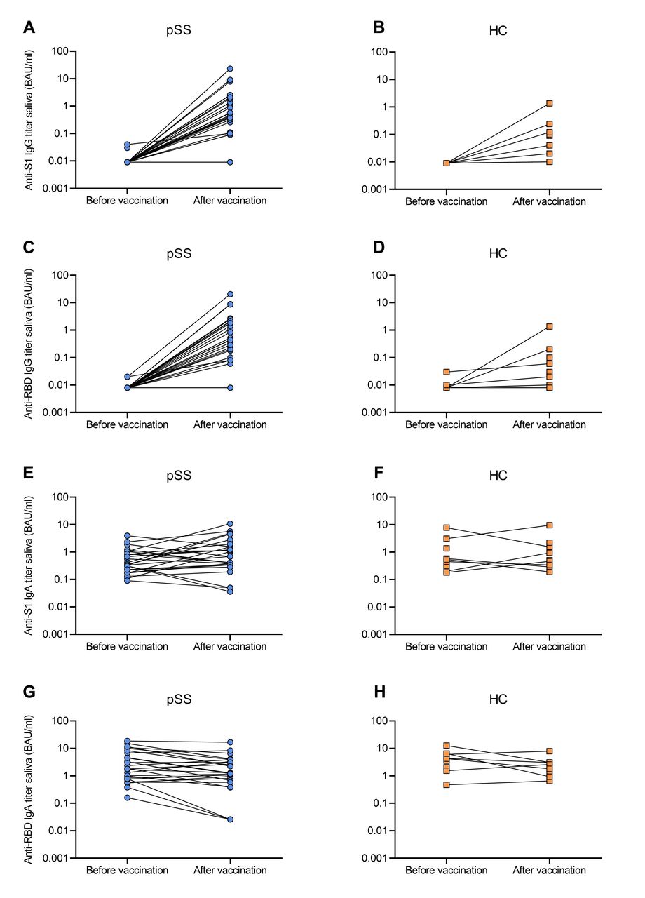
Change in saliva in (A) anti-S1 IgG levels in patients with pSS (B) anti-S1 IgG levels in HC (C) anti-RBD IgG levels in patients with pSS (D) anti-RBD IgG levels in HC (E) anti-S1 IgA levels in patients with pSS (F) anti-S1 IgA levels in HC (G) anti-RBD IgA levels in patients with pSS (H) anti-RBD IgA levels in HC. HC, healthy controls; pSS, primary Sjögren’s syndrome; RBD, receptor binding domain; S1, spike 1.
Adverse events
No serious AEs (SAEs) occurred in this study. Taking all vaccine types together, frequencies of (systemic) AEs were comparable between patients with pSS and HC (figure 4 and table 2). After the second vaccination, arthralgia and myalgia were more severe in patients with pSS compared with HC (p=0.024 and p=0.016, respectively). Analyses of AEs split for the separate vaccine types also revealed no significant differences in frequencies of AEs (online supplemental figure 4, table 2). The frequency of systemic AEs in the pSS and HC groups was also comparable to the Lareb general population cohort16 (online supplemental figure 4).
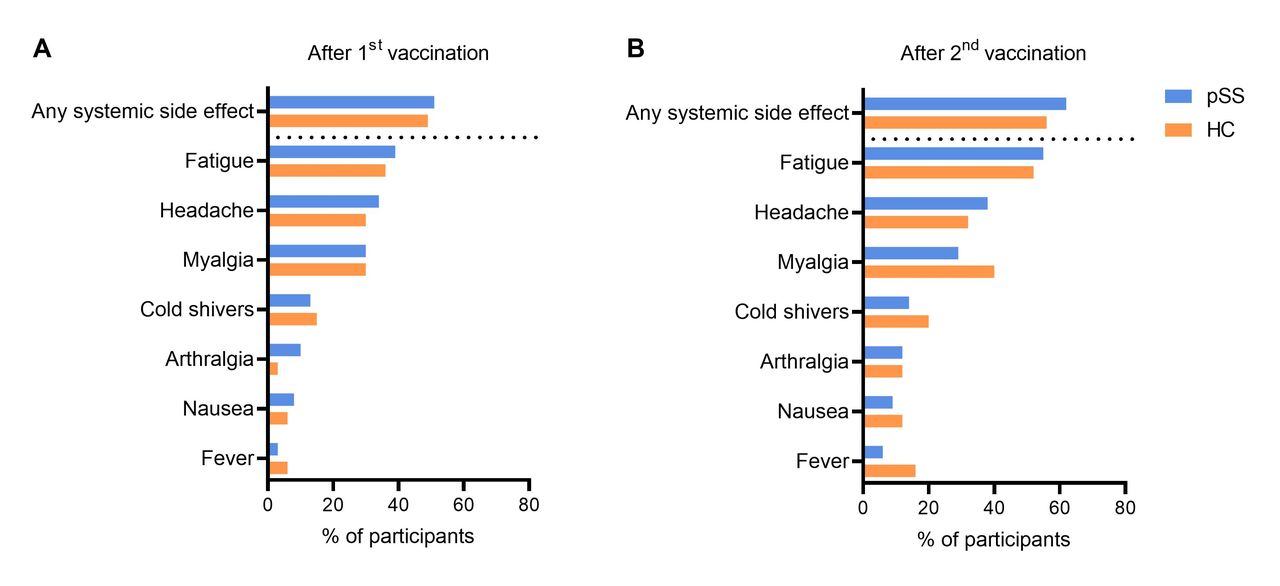
Systemic side effects after the 1st and 2nd vaccination in patients with pSS and HC. HC, healthy controls; pSS, primary Sjögren’s syndrome.
Frequency of (systemic) adverse events (AEs) for the total group and split for Pfizer-BioNtech, AstraZeneca and Moderna
Disease activity
For patients with pSS, patient-reported disease activity measured with ESSPRI did not change from baseline (median 6, IQR 5–7) to 28 days after the second vaccination (first for Janssen) (6, IQR 4–7, p=0.16) (table 3). Furthermore, total serum IgG levels did not change after vaccination. In total, 36 patients with pSS had an available ESSDAI within 6 months after vaccination (recorded until December 2021). Median ESSDAI did not change from baseline (3, IQR 1–4) to after vaccination (2, IQR 0–5, p=0.88). Disease activity parameters also remained stable in the Pfizer-BioNtech, AstraZeneca and Moderna groups separately (online supplemental table 5).
Change in patient-reported and systemic disease activity and IgG levels after vaccination for all patients with pSS
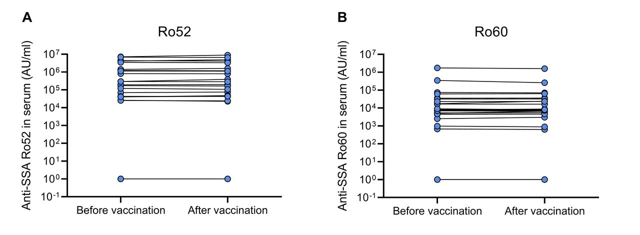
In 26 patients with pSS, anti-SSA antibodies were measured in blood samples before and after vaccination. No changes were seen in anti-Ro52 and anti-Ro60 antibody levels (p=0.65 and p=0.58, respectively) (figure 5).
(A) Anti-SSA Ro52 and (B) Ro60 antibody levels before and after vaccination in patients with pSS. pSS, primary Sjögren’s syndrome.
Discussion
In this prospective longitudinal study, patients with pSS generated similar anti-SARS-CoV-2 antibody levels following COVID-19 vaccination compared with HC, suggesting that COVID-19 vaccination is effective in patients with pSS. Spike-specific T-cell response also seems similar between patients with pSS and HC. Besides an increase in systemic spike-specific antibodies, increases in IgG antibodies, but not IgA, were detected in saliva. None of the participants experienced SAEs, and frequencies of AEs were comparable between patients with pSS and HC. Furthermore, no worsening was observed in patient-reported and systemic disease activity or auto-antibody levels following vaccination in patients with pSS.
To the best of our knowledge, this is the first study reporting on COVID-19 vaccination response in patients with pSS without immunosuppressive treatment. Most studies included patients with various rheumatic diseases who were treated with immunosuppressive drugs, which contained only few patients with pSS, and results were not given separately for patients with pSS.3–5 21 Prevailing evidence shows that the most important factors associated with attenuated spike-specific antibody response are specific immunosuppressive medications, such as anti-CD20 therapy.3–5 21 In our study, no significant differences in spike-specific antibody levels between patients with pSS and HC were found, which is in line with most other studies with untreated rheumatic disease patients.5 22 One prospective cohort study showed that untreated rheumatic disease patients obtained similar seroconversion rates after the second vaccination compared with HC (23/26 (88%) vs 38/40 (95%)).5 However, one study in 24 untreated patients with various rheumatic diseases showed a delayed and reduced spike-specific antibody response.23 A cohort study of 126 patients with systemic lupus erythematosus found, similar to our study, no associations of SARS-CoV-2 antibody response with hydroxychloroquine use or baseline disease activity scores, but higher baseline total IgG levels were associated with higher anti-SARS-CoV-2 antibody levels, which was in contrast to our study.24 Although anti-spike IgG levels correlate with virus neutralising activity,25 the threshold level that is required to provide protection against COVID-19 remains unclear, especially against new variants. In addition to antibodies, T-cells also play a key role in viral clearance and limiting disease.26 We observed that the majority of patients with pSS were also T-cell responder, indicating that the two arms of the immune system are activated in patients with pSS following vaccination.
Although we hypothesised that patients with pSS may have an increased humoral response following COVID-19 vaccination compared with HC as a consequence of (TLR-7/type-1 IFN mediated) B-cell hyperactivity, we did not find this in our study. This is in contrast to influenza vaccination studies in pSS, which demonstrated that untreated patients with pSS acquired higher influenza-specific antibody responses following H1N1 vaccination, compared with HC.7 8 Furthermore, an increase in anti-SSA auto-antibody levels was observed following vaccination.7 8 Patients with pSS treated with hydroxychloroquine generated similar levels of influenza-specific antibodies as HC, which could be due to the modulating effect on the TLR-7 pathway.7 We did not find an effect of hydroxychloroquine use on anti-SARS-CoV-2 antibody levels among patients with pSS. The influenza vaccines used in these studies were a squalene-adjuvanted inactivated split virion H1N1 vaccine (Pandemrix),8 and a non-adjuvanted inactivated split virion H1N1 vaccine (Fluarix) containing antigens of several influenza virus strains.7 The fact that we did not observe higher SARS-CoV-2 specific antibody responses in patients with pSS than HC, and no increase in auto-antibodies, may be explained by the different working mechanism of COVID-19 vaccines, that is, not containing adjuvants, and including only a single specific virus gene encoding for spike protein. This difference might also be due to the presence of pre-existing immunity against influenza as the result of prior infections and vaccinations, whereas no anti-spike immune memory was present in our study population at the beginning of this study.
Our study included untreated patients with pSS, beyond hydroxychloroquine, and the majority (78%) had a low ESSDAI score. We cannot rule out the possibility that patients with high systemic disease activity respond differently to COVID-19 vaccination than patients with low systemic disease activity. However, in our regression analyses we did not observe an effect of ESSDAI on anti-SARS-CoV-2 antibody levels. For patients with pSS treated with immunosuppressives, it is still important to take into account the effect of their medication on COVID-19 vaccination response.
Because salivary glands are involved in the disease process of pSS, anti-SARS-CoV-2 antibody levels in saliva samples are of particular interest. Unfortunately, groups were too small to statistically compare antibody levels between patients with pSS and HC. However, an increase in anti-S1 and anti-RBD IgG levels was observed in both groups and a significant correlation was seen with serological anti-SARS-CoV-2 antibody levels. This is not unexpected since most salivary IgG derives from serum antibodies via gingival crevices. We observed no increase in anti-S1 and anti-RBD IgA levels. In contrast, some studies did show a mucosal IgA response,27 28 although this was often weaker than the mucosal IgG and systemic antibody response. However, some other studies also reported no mucosal IgA response following vaccination.29 30
In addition to immunogenicity, safety of COVID-19 vaccination was assessed. Frequencies of (systemic) AEs were comparable between patients with pSS and HC. Sjögren’s disease activity, measured by patient-reported, systemic and serological parameters, did not worsen following vaccination. Postvaccination ESSDAI scores were only available for approximately half of patients with pSS, but also appeared to remain stable. These findings are similar to other studies in rheumatic disease patients reporting on AEs and/or disease activity following COVID-19 vaccination.3 31 32 One study in 505 patients reported no difference in systemic AEs between patients with rheumatic diseases (44%) and HC (40%).31 Another study in 5121 patients with rheumatic diseases showed that only a small number (4.4%) experienced a disease flare following vaccination, which were mostly mild or moderate.33 Most other studies also did not find an increase in disease activity following COVID-19 vaccination in rheumatic disease patients.3 24 34
A main limitation of this study is that four different vaccine types were included, which were administered according to the national vaccination programme. This led to an unequal distribution of participants within the distinct vaccination groups. Relatively more participants in the HC group received the Moderna or Janssen vaccine, compared with patients with pSS. Other limitations are single-centre recruitment and a relatively small sample size, especially for certain vaccine types. Because of the small HC group, not all subgroup measurements could be statistically compared with patients with pSS.
In conclusion, COVID-19 vaccination led to similar anti-SARS-CoV-2 antibody levels and T-cell responses in patients with pSS without immunosuppressives compared with HC, providing evidence that COVID-19 vaccination is effective in patients with pSS. Furthermore, side effects were comparable between patients with pSS and HC, and no increase in disease activity was seen, indicating that COVID-19 vaccination is safe for patients with pSS. Our findings are an important and reassuring message for patients with pSS and provide arguments that patients with pSS may also benefit from future booster vaccinations.
Data availability statement
No data are available. Currently there are no plans to share additional data beyond what is included in this article.
Ethics statements
Patient consent for publication
Ethics approval
This study involves human participants and ethical approval was obtained from the UMCG institutional review board (METc 2021.084). All participants provided informed consent according to the Declaration of Helsinki. Participants gave informed consent to participate in the study before taking part.
Acknowledgments
We thank all patients for participation in our study. We thank Martine Tax and Joan Lommen from Labonovum for their collaboration on the anti-SARS-CoV-2 antibody assay in the finger prick samples. We thank the National Institute of Public Health and Environment (RIVM) for their collaboration on the anti-SARS-CoV-2 antibody assays in the serum samples. We thank Idil Esen and Elisabeth Raveling-Eelsing for their help with the ELIspot assays for spike-specific T-cell response. We thank Ellis Herder and Janita Bulthuis for their help with the logistics of this study.
References
Supplementary materials
Supplementary Data
This web only file has been produced by the BMJ Publishing Group from an electronic file supplied by the author(s) and has not been edited for content.
Footnotes
Twitter @sleenyannick
GMV and LdW contributed equally.
Contributors GV, LdW, DvB, FK and HB contributed to the study design and developed the study protocol. GV, H-MH and LdW recruited patients for inclusion in this study. GV, LdW, H-MH, YvS, AV, JHT, DAD, MvdH and HB contributed to collection of the data. GV, LdW, SA, YvS, DAD, AV, DB, FK and HB contributed to analysis and interpretation of the data. LdW wrote the first draft of the manuscript. GV is guarantor for this study and accepts full responsibility for the overall content of this study. All authors contributed to manuscript revision, read and approved the submitted version.
Funding This study was funded by the Dutch Sjögren’s Patients Association (NVSP; Nationale Vereniging Sjögrenpatiënten) and AstraZeneca BV. The funders had no role in study design, data collection, data analysis, data interpretation or writing of the report.
Competing interests HB: reports grants and personal fees from Bristol Myers Squibb and Roche, and personal fees from Novartis, Medimmune, personal fees and Union Chimique Belge, outside the submitted work.
Provenance and peer review Not commissioned; externally peer reviewed.
Supplemental material This content has been supplied by the author(s). It has not been vetted by BMJ Publishing Group Limited (BMJ) and may not have been peer-reviewed. Any opinions or recommendations discussed are solely those of the author(s) and are not endorsed by BMJ. BMJ disclaims all liability and responsibility arising from any reliance placed on the content. Where the content includes any translated material, BMJ does not warrant the accuracy and reliability of the translations (including but not limited to local regulations, clinical guidelines, terminology, drug names and drug dosages), and is not responsible for any error and/or omissions arising from translation and adaptation or otherwise.