Article Text

Abstract
Rheumatoid arthritis (RA) and psoriatic arthritis (PsA) have key differences in clinical presentation, radiographic findings, comorbidities and pathogenesis to distinguish between these common forms of chronic inflammatory arthritis. Joint involvement is typically, but not always, asymmetric in PsA, while it is predominantly symmetric in RA. Bone erosions, without new bone growth, and cervical spine involvement are distinctive of RA, while axial spine involvement, psoriasis and nail dystrophy are distinctive of PsA. Patients with PsA typically have seronegative test findings for rheumatoid factor (RF) and cyclic citrullinated peptide (CCP) antibodies, while approximately 80% of patients with RA have positive findings for RF and CCP antibodies. Although there is overlap in the pathogenesis of PsA and RA, differences are also present that affect the efficacy of treatment. In PsA, levels of interleukin (IL)-1β, IL-6, IL-17, IL-22, IL-23, interferon-γ and tumour necrosis factor-α (TNF-α) are elevated, and in RA, levels of IL-1, IL-6, IL-22, IL-33, TNF-α, chemokine ligand 11 and chemokine C-X-C motif ligand 13 are elevated. Differences in the pathogenesis of RA and PsA translate into some variances in the specificity and efficacy of therapies.
- psoriatic arthritis
- rheumatoid arthritis
- inflammatory disease
This is an Open Access article distributed in accordance with the Creative Commons Attribution Non Commercial (CC BY-NC 4.0) license, which permits others to distribute, remix, adapt, build upon this work non-commercially, and license their derivative works on different terms, provided the original work is properly cited and the use is non-commercial. See: http://creativecommons.org/licenses/by-nc/4.0/
Statistics from Altmetric.com
Key messages
What is already known about this subject?
Rheumatoid arthritis (RA) and psoriatic arthritis (PsA) are both common chronic inflammatory diseases, and differentiation of these conditions can be challenging.
What does this study add?
We review key differences in clinical, serological, radiographic and pathogenic features of RA and PsA, and provide practical guidance for achieving a correct diagnosis.
How might this impact on clinical practice?
Accurate diagnosis of RA and PsA is important because differences in underlying pathogenesis and response to therapy translate into substantially different clinical outcomes.
Introduction
Rheumatoid arthritis (RA) and psoriatic arthritis (PsA) are common chronic inflammatory diseases; both are characterised by pain and swelling in the joints and have significant systemic manifestations.1–3 If not diagnosed and treated early, both can lead to joint destruction with loss of function. For this reason, early diagnosis is important to determine therapeutic strategies that will optimise clinical and radiographic outcomes.4 Differentiating between RA and PsA can be clinically challenging because there are many similarities in their clinical presentation and manifestations. Both also have similarities with other inflammatory diseases5 and association with coprevalent forms of arthritis, such as gout and secondary osteoarthritis.6–8
In the last 20 years, biologic disease-modifying antirheumatic drugs, many with differing mechanisms of action, have become available. These medications target the disease processes that drive inflammation and joint damage in RA and PsA. However, depending on the molecule, therapeutic selection is complicated by variable efficacy between patients with RA and the manifestations of psoriatic disease (eg, enthesitis or skin psoriasis).
To help practitioners determine an accurate diagnosis, key differences in clinical, serological, radiographic and pathogenic features of RA and PsA are reviewed. The different therapeutic classes that can be of benefit in each disease are discussed in depth.
Differences of RA and PsA
RA is an autoimmune systemic inflammatory disease characterised by synovitis, bony erosions and cartilage damage.9 PsA is a heterogeneous autoimmune systemic disease with diverse clinical and radiographic manifestations.9 The presence of psoriasis precedes the development of PsA in 85% of patients, and PsA typically develops about 10 years after the onset of psoriasis.10 Other common clinical features of PsA include synovitis with subsequent osteolysis and/or joint fusion of peripheral joints, axial involvement, sacroiliitis, and extra-articular manifestations, including nail dystrophy, enthesitis and dactylitis; not all are present in every patient. Key clinical, serological and radiographic differences between RA and PsA are summarised in table 1.
Clinical, serological and radiographic characteristics of PsA and RA
Epidemiological characteristics of RA and PsA
RA is more common than PsA, affecting more than 1 million in the USA.11 PsA affects roughly half a million people in the USA12 and approximately 30% of patients with psoriasis.13 Worldwide prevalence estimates for RA and PsA are variable. In many populations, RA prevalence is estimated to be 0.5%–1.0%; however, prevalence is much higher in Native American Indian populations (~5% to 7%) but lower in China and Japan (~0.2% to 0.3%).14–19 PsA prevalence estimates in the USA and Europe range from 0.1% to 0.4%, whereas in Japan, PsA prevalence is lower.12 20 21 This variability in prevalence suggests that both environmental and genetic factors affect risk for disease.
Pathogenesis of RA and PsA
A combination of genetic factors and environmental triggers is thought to elicit autoimmune inflammatory responses in both RA and PsA. The pathogenesis of RA and PsA is not completely understood. In addition to the known association with human leucocyte antigen (HLA)-DR4 in RA, one theory is the development of lung inflammation, typically prior to joint symptoms, with production of antibodies to citrullinated protein antigens that mediate pathogenesis.22 23 Gut dysbiosis has been linked with the pathogenesis of PsA.24 While there is some overlap in the development of inflammation in PsA and RA, some important differences are evident.25 For example, in both PsA and RA, HLA alleles have been shown to affect disease susceptibility and severity; however, the primary genotypes associated with each disease are different. In PsA, HLA-B27 is associated with the development of enthesitis and symmetric sacroiliitis, and HLA-B08 is associated with joint fusion, asymmetric sacroiliitis and dactylitis.26 In RA, HLA-DRB1 alleles are associated with disease susceptibility and severity in patients who have positive findings for rheumatoid factor (RF) and cyclic citrullinated peptide (CCP) antibodies.27
In both PsA and RA, inflammatory responses are characterised by the increased production of proinflammatory molecules that act in synergy to propagate chronic inflammation (figure 1).25 28 29 In PsA, activated T cells and macrophages induce production of inflammatory chemokines and cytokines, including interleukin (IL)-17, IL-23, IL-22, IL-1β, IL-6, interferon-γ and tumour necrosis factor-α (TNF-α).29 30 Specifically, elevated levels of IL-17 + CD8+ T cells have been observed in the joints of patients with PsA but not in those with RA.31 In addition, recent studies have implicated increased expression of IL-9 and its receptor (IL-9R) in the promotion of pathological T-cell growth and subclinical gut inflammation.32 33

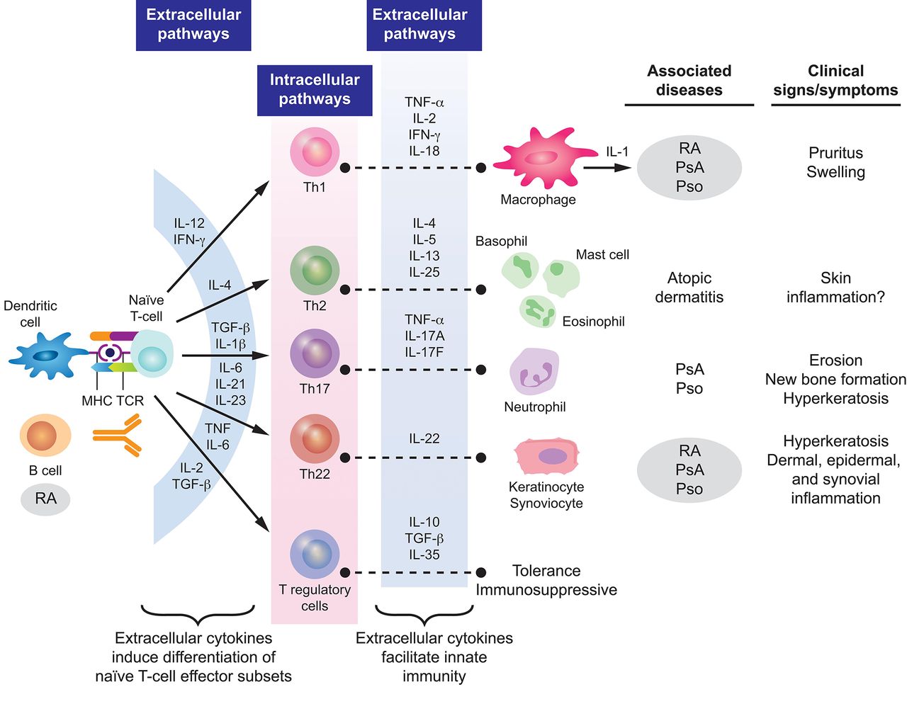
Pathogenesisof PsA and RA. Reprinted from Coates et al 29 and Perera et al.28 IFN, interferon; IL, interleukin; MHC, major histocompatibility complex; PsA, psoriatic arthritis; Pso, psoriasis; RA, rheumatoid arthritis; TCR, T cell receptor; TGF-β, transforming growth factor-β; Th, T helper; TNF-α, tumour necrosis factor-α.
Key cytokines identified as being associated with joint damage and radiographic progression of RA include TNF-α, IL-6, IL-1, IL-22, IL-33, chemokine ligand 11 and chemokine C-X-C motif ligand 13.25 Certain cytokines and chemokines are present at higher levels in patients with polyarticular versus oligoarticular arthritis, suggesting that they could be markers of disease severity and/or progression.25 In addition, it has been hypothesised that the onset of RA may be triggered in some patients by autoimmune responses (eg, elevated CCP levels) in the lungs. Smoking, infections or air pollution exposure may trigger local inflammation in the lungs, promoting protein citrullination and stimulating CCP production.34 35
Clinical characteristics of RA and PsA
For RA, the American College of Rheumatology (ACR)/European League Against Rheumatism classification criteria were designed for patient characterisation and use in clinical trials.36 The key clinical characteristic is the confirmation of definite, persistent, clinical synovitis in at least one joint. The criteria include the number of joints involved, duration of symptoms, and the demonstration of serological markers and an elevated acute-phase reactant. For PsA, the Classification Criteria for Psoriatic Arthritis help categorise patients with inflammatory articular disease for clinical trials.37 Key clinical characteristics include a personal or family history of psoriasis, psoriatic nail dystrophy and dactylitis. Neither classification criteria should be confused as diagnostic criteria.
Joint involvement is predominantly symmetric in RA and often, but not always, asymmetric in PsA.25 38 39 In both RA and PsA, most patients have polyarthritis (≥5 involved joints), although joint involvement can be oligoarticular or polyarticular.5 25 26 39 40 Monoarticular disease is less common in PsA; however, 5%–10% of patients may present with isolated distal joint involvement.5 In PsA, prognosis worsens and symmetry of joint involvement tends to increase as the number of affected joints increases.5 41
Typically, RA affects the shoulder, elbow, wrist, metacarpophalangeal, proximal interphalangeal, hip, knee, ankle and metatarsophalangeal joints. In PsA, the distal interphalangeal joints of the hands and feet, large joints of the lower extremities, the axial spine and sacroiliac joints are commonly affected; the metacarpophalangeal and metatarsophalangeal joints and wrist can be involved as well. PsA, rather than RA, is included in the spectrum of spondyloarthritis as PsA can affect the axial skeleton (eg, sacroiliac joints and spine).26 38 It is estimated that up to 50% of patients with PsA experience inflammation in the axial skeleton42; axial involvement can be a differentiating feature of PsA because it is not present in RA other than cervical spine involvement, which has been reported in up to 80% of patients with RA.43
Although TNF-α induces angiogenesis in both RA and PsA,44 differences in synovial vascularity can help differentiate the diseases. Both RA and PsA exhibit proliferation of endothelial cells, but topological differences in endothelial cells suggest differing pathological features.45 Among patients with knee synovitis, straight, branching vessels are observed in RA, and predominantly tortuous, bushy vessels are observed in PsA.46 These physiological differences may be caused by varied patterns of synovial cytokine expression as significantly higher levels of IL-1β, IL-2, IL-10 and IFN-γ are found in PsA synovial explants compared with RA synovial explants.47
Enthesitis (inflammation of entheses at sites where ligaments or tendons insert into the bone) occurs in 35% of patients with PsA48 but is uncommon in patients with RA.39 Enthesitis is proposed to have a key role in the pathogenesis of PsA,49 and the presence of enthesitis can be particularly valuable in differentiating PsA from RA. The most common locations of enthesitis in patients with PsA are the insertion sites of the plantar fascia, the Achilles tendon and ligamentous attachments of the knee.50 In rare cases, enthesitis may be the only manifestation of PsA. Signs and symptoms of enthesitis can be non-specific and difficult to distinguish from other inflammatory conditions, particularly fibromyalgia, in the absence of other manifestations common to PsA.40
Dactylitis (inflammation of an entire digit) is a common manifestation of PsA that affects up to 50% of patients, compared with approximately 5% of patients with RA.5 39 Dactylitis may also occur in patients with other forms of spondyloarthritis, such as ankylosing spondylitis and reactive arthritis, but also occasionally in gout and sarcoidosis.5 26 51 However, dactylitis is a fairly specific sign of PsA that can aid in the differential diagnosis, especially when it is present along with enthesitis.
Nail dystrophy, which is characterised by onycholysis, pitting and hyperkeratosis, is an important clinical manifestation of PsA.5 In a prospective study characterising the clinical presentation of early PsA, 67% of patients had nail dystrophy; the incidence was higher (80%) in patients with distal interphalangeal joint involvement.52 The increased incidence of nail disease in patients with distal interphalangeal joint involvement has been attributed to the topographic association between the extensor tendon enthesis and the nail.53 Thus, the presence of nail lesions can be especially helpful in differentiating PsA from other forms of spondyloarthritis, RA, gout and osteoarthritis.
Ocular disorders are common extra-articular manifestations of RA and PsA.54 55 The most common ocular manifestation of RA is keratoconjunctivitis sicca, which affects approximately 18% of patients; other common eye disorders in patients with RA include episcleritis (5%) and scleritis (2%).56 Uveitis occurs in PsA, affecting approximately 7% of patients and is more common in women than men; the incidence is far higher in patients with axial disease and HLA-B27 positivity.54 57–59 In patients with PsA or inflammatory bowel disease, uveitis is typically bilateral with an insidious onset, affecting the anterior and intermediate layers of the eye.54
Cutaneous manifestations are common in both RA and PsA. The most common cutaneous features of RA include rheumatoid nodules, vasculitic skin lesions and granulomatous dermatoses.60–62 Current psoriasis is an important clinical finding in a patient with arthritis. Skin disease precedes the development of joint symptoms in 84% of patients with PsA, and up to 96% of patients with PsA have either current or previous psoriasis or family history of psoriasis.63 Compared with patients with psoriasis but without PsA, patients with PsA have more extensive psoriasis and higher rates of pustular and inverse psoriasis.64 In addition, patients with scalp psoriasis, inverse psoriasis and nail dystrophy are at increased risk for PsA.65 It is important to remember that manifestations of psoriasis are not always overt, and patients with seronegative inflammatory arthritis should be evaluated for scalp, inverse, genital, palmoplantar and nail psoriasis. Furthermore, inverse psoriasis is traditionally under-recognised, and recent findings indicate a greater prevalence of inverse psoriasis than commonly appreciated.66 With such a thorough physical examination, many patients with ‘seronegative’ RA are correctly identified as actually having PsA.
Serological features of RA and PsA
RA is a seropositive arthropathy, with approximately 80% of patients having a positive test result for RF or CCP antibodies.39 67 CCP antibodies are a more specific marker for RA than RF, but both biomarkers are considered to be distinct and complementary predictors of disability and joint erosion.68 69
In contrast, PsA is a seronegative inflammatory arthropathy. RF and CCP are absent in most patients with PsA, and if patients do have positive test findings for RF or CCP, the titres are usually low.2 39 67 70 In a study comparing patients with RA or PsA and controls, the mean RF and anti-CCP titre values were substantially higher in patients with RA compared with PsA (RF titre: 56 vs 11 U/mL; anti-CCP titre: 14 vs 2 U/mL).67 Titres in patients with PsA were similar to values in controls.67 Although the presence of serum RF or CCP antibodies is generally not used to exclude diagnosis of non-rheumatic diseases (eg, fungal infections), data suggest that at anti-CCP titre values ≥11.6 U/mL, it is highly probable that patients have RA rather than PsA.67 In both patients with RA and PsA, the presence of anti-CCP antibodies is associated with bone destruction, suggesting that the osteocatabolic effect of anti-CCP antibodies is not found only in RA as previously thought.71
C reactive protein (CRP) and erythrocyte sedimentation rate (ESR) are markers of acute-phase inflammatory responses in patients with RA and PsA, but more so in patients with RA.36 70 In a systematic literature review of RA disease activity parameters, ESR and CRP levels were a significant predictor of radiographic progression in most studies.72 The acute-phase response is correlated with synovial inflammation, radiographic disease progression and erosive joint damage.25 On average, patients with PsA have significantly lower CRP and ESR levels than patients with RA.73 However, elevated ESR and CRP levels are significantly correlated with the number of swollen joints and with ultrasound abnormalities in PsA.73–75 ESR, in particular, is considered to be one of the best predictors of damage progression in PsA. It has been suggested that a low ESR is protective, while rates >15 mm/hour are associated with increased risk for mortality.70 Changes in ESR and CRP correspond with clinical outcomes and can be used to track response to treatment.73
Increased ESR and CRP levels are markers of inflammation, but not necessarily just in RA.76 Other rheumatological diseases associated with elevated ESR and CRP levels include polymyalgia rheumatica, Sjögren’s syndrome and ankylosing spondylitis.76
Radiographic features of RA and PsA
Use of imaging can provide important information to help practitioners identify and differentiate between types of inflammatory arthritis. Conventional radiography can be used to identify juxta-articular bony proliferations, which can be used to help discern PsA from other types of inflammatory arthritis, and to visualise osteodestructive lesions characteristic of RA.77 Other characteristic radiographic changes observed in patients with severe PsA (including arthritis mutilans) are bone resorption, pencil-in-cup deformities and ankylosis.26 However, conventional radiographs are not as sensitive as ultrasound or MRI for detection of bone erosions and may not help clinicians detect soft-tissue changes well.78 Additionally, in our experience, radiologists will often report changes consistent with osteoarthritis in patients with PsA, and it is necessary for rheumatologists to interpret these findings in the proper clinical context.
Ultrasound imaging can be used to identify characteristic features of inflammatory arthritis, including enthesitis, cortical bone erosions, cartilage lesions, synovitis and tenosynovitis.78 Bone erosions are an important diagnostic criterion of RA that can be identified based on intra-articular discontinuity of the bone surface.78 79 Ultrasound evaluations of bone erosions are more reliable for joints that are easily accessible (eg, small joints of hands or feet) than for carpal or tarsal bones, which cannot be viewed circumferentially.78
Interestingly, while clinical signs of enthesitis are a hallmark of PsA, the presence of enthesitis on ultrasound cannot always help clinicians distinguish PsA from RA.80 In an evaluation of the Leeds Enthesitis Index, greyscale and power Doppler techniques could not help distinguish between PsA and RA based on features of enthesitis, possibly because ultrasound imaging cannot visualise bone oedema, which may cause entheseal tenderness, or because of a higher prevalence of juxta-articular inflammation in RA.80 However, it has been observed that central slip enthesitis at the proximal interphalangeal joints is present on ultrasound in about 25% of patients with PsA but absent in patients with RA, potentially serving as a distinguishing feature of PsA versus RA in some patients.1
Ultrasound imaging can be used to identify characteristic joint involvement that distinguishes PsA from RA. For example, distal interphalangeal joint changes are found almost exclusively in patients with PsA compared with those with RA,81 and dactylitis due to flexor tenosynovitis is characteristic of PsA.81–83 While overall prevalence of synovitis is higher in RA (~90% of affected joints) than in PsA (~60% of affected joints), peritendon extensor digitorum tendon inflammation is significantly more common in early PsA than in early RA (54.1% vs 2.5%).1 It can be difficult to differentiate between types of inflammatory arthritis based on bone changes observed on ultrasound; however, cortical irregularity from periosteal new bone formation is suggestive of PsA.84
MRI can be used to identify joint synovitis, bone and joint erosions, bone marrow oedema, spondylitis, periarticular inflammation, active enthesitis, nail disease and periostitis.85 86 The location of bone marrow oedema can help differentiate between PsA (near entheses) and RA (near capsular attachments), and diaphyseal bone marrow oedema is a characteristic feature of early PsA.87 As with ultrasound, on MRI, distal interphalangeal joint erosions and hypertrophic bone changes are characteristic findings in PsA that may help differentiate it from RA. Tenosynovitis is another common MRI observation in inflammatory arthritis; in RA, tenosynovitis is most common in the hands and wrists, whereas in PsA soft-tissue inflammation around the tendon sheath is characteristic of dactylitis.86 MRI is also useful for identifying patients that will develop onychopathy, a characteristic of psoriatic nail dystrophy.85
Comorbidities occurring in RA and PsA
Differences in patient comorbidities may help clinicians differentiate between RA and PsA (table 2). Overall, comorbidity burden may be higher in RA than in PsA, but both diseases are similarly associated with increased risk for comorbidities linked to systemic inflammation (eg, cardiovascular disease).29 88 Han and colleagues89 found that patients with RA and PsA had similarly increased prevalence ratios of ischaemic heart disease, atherosclerosis, peripheral vascular disease, congestive heart failure, cerebrovascular disease, hyperlipidaemia and hypertension compared with healthy controls. However, registry data suggest that the rates of obesity, diabetes mellitus and metabolic syndrome are significantly higher in patients with PsA compared with those with RA.90 Notably, most patients with PsA are overweight or obese.90 Cardiometabolic comorbidities of PsA are associated with higher levels of systemic inflammation and increased disease severity.91 92 In addition, psoriatic skin lesions are associated with an increased risk for cardiovascular disease and mortality.93 94 Of interest, PsA is an independent predictor of non-alcoholic fatty liver disease (NAFLD) in patients with psoriasis, while in patients with RA, the rates of NAFLD are similar to those observed in the general population.29 95
Summary of differences in common comorbidities associated with PsA and RA29 88 90 182–185
Treatment options for RA and PsA
Because of the differences in disease pathogenesis, clinical manifestations and response to therapy between RA and PsA, treatment strategies may differ. Table 3 provides a summary of current Food and Drug Administration (FDA)-approved treatments for RA and PsA. Agents targeting more upstream factors (eg, TNF-α) are effective in both PsA and RA, while agents targeting more downstream cytokines are more disease-specific, demonstrating significant efficacy in either RA (eg, IL-6) or PsA (eg, IL-17A), but not in both diseases.
FDA-approved therapies for PsA or RA
Conventional synthetic disease-modifying antirheumatic drugs
Methotrexate is the most frequently used conventional synthetic disease-modifying antirheumatic drug (csDMARD) for the treatment of RA, and its efficacy is well-established in this patient population.96 Data on the use of methotrexate in PsA are more mixed, and it is believed that, generally, methotrexate is not effective for every clinical domain affected by PsA.97 No benefit was observed with methotrexate compared with placebo in a randomised, placebo-controlled trial in PsA,98 but this trial has been heavily criticised as it was underpowered and used a suboptimal dose of methotrexate. However, a benefit of methotrexate monotherapy was observed in the ‘tight control of psoriatic arthritis’ study.99 Although methotrexate has not been studied in large-scale controlled trials in PsA and the studies that have been conducted using methotrexate have likely underestimated its efficacy because of limitations of study designs, real-world data indicate that the efficacy of methotrexate in PsA may be comparable with that in RA.96 100 In addition, methotrexate is associated with reductions in disease activity and improvements in health-related quality of life in patients with PsA and RA.96 While leflunomide has shown efficacy in patients with PsA compared with placebo, it is only FDA-approved for the treatment of patients with RA in the USA.101
Biologic disease-modifying antirheumatic drugs
TNF-α inhibitors
TNF-α is a proinflammatory cytokine that is overexpressed in the synovium of patients with inflammatory arthritis. Currently, five TNF-α inhibitors (etanercept, infliximab, adalimumab, golimumab and certolizumab pegol) have been FDA-approved for the treatment of both RA and PsA in the USA.
In RA, a consequence of TNF-α inhibition, in addition to inhibiting systemic inflammation, is to inhibit bone erosion and reduce structural damage by inhibiting osteoclast formation. The ability of TNF-α inhibitors to block induction of matrix metalloproteinases and aggrecanases has been postulated to reduce destruction of the cartilage matrix.102 103 Across clinical studies in RA, the five available TNF-α inhibitors have provided strikingly similar improvements in ACR20, ACR50 and ACR70 response rates, reductions in structural damage, and improvements in quality of life.104–111
In PsA, TNF-α inhibitors reduce synoviocyte hyperproliferation and decrease T-cell and macrophage infiltration, reducing synovial thickness.112–115 Similar to their actions in RA, TNF-α inhibitors modulate angiogenesis and osteoclastogenesis and reduce synovitis in PsA.113 114 116 They have also been shown to improve signs and symptoms of enthesitis, dactylitis, axial involvement and skin disease, and evidence suggests that TNF-α inhibition slows radiographic disease progression.117–119
IL-17A inhibitors
IL-17A is a proinflammatory effector cytokine produced by T helper (Th)17 cells, macrophages, mast cells, dendritic cells, natural killer cells and CD8+ T cells.120–125 Synovial fluid levels of IL-17A are increased in patients with PsA, and studies have shown that this cytokine plays a role in pathogenic angiogenesis, osteoclastogenesis and fibrogenesis.31 126–128 IL-17A interactions with synovial-like fibroblasts, osteoblasts and osteoclast precursors promote chronic inflammation and bone changes that contribute to joint damage in PsA.129–131 Like TNF-α, IL-17 can also induce osteoclastogenesis and upregulate matrix metalloproteinases and other proinflammatory cytokines.31 In patients with PsA, the presence of IL-17-producing T cells is associated with disease activity measures (ie, CRP and ESR) and radiographic erosion.31
The IL-17A inhibitors secukinumab and ixekizumab have been FDA-approved for the treatment of active PsA. In clinical studies, treatment with secukinumab and ixekizumab improved signs and symptoms of disease, inhibited radiographic progression of joint damage, and improved physical functioning and quality of life in patients with active PsA.132–134 In addition, secukinumab and ixekizumab were efficacious in patients who were naïve to TNF-α inhibitors, had an inadequate response to TNF-α inhibitors, or stopped treatment because of safety or tolerability reasons.133 135 136
Enhanced expression of IL-17A is a characteristic feature in the joints of patients with PsA but not RA. Consistent with this finding, clinical studies of IL-17A inhibitors in RA have shown that these agents have limited therapeutic efficacy.137–139
IL-12 and IL-23 dual inhibitor
IL-12 and IL-23 are signature cytokines involved in the pathogenesis of psoriasis and PsA. IL-12 stimulates Th1 cells and is produced by monocytes and macrophages in response to inflammation, and IL-23 is an upstream cytokine produced predominantly by myeloid dendritic cells to regulate Th17 cell function.140–144 IL-12 promotes continued Th1 differentiation, inflammation and activation of natural killer cells, and IL-23 is involved in the pathogenic processes related to osteoclastogenesis and bone erosion.140 145–147
The IL-12/23p40 subunit inhibitor, ustekinumab, blocks Th1 and Th17 differentiation and downstream production of IL-17. In phase III clinical trials, ustekinumab significantly reduced radiographic progression of joint damage and improved signs and symptoms of disease in patients with active PsA.148 149 Ustekinumab has not demonstrated efficacy in patients with RA.150 An IL-23-specific inhibitor, guselkumab, was also not efficacious in patients with RA151; however, preliminary data of this agent in patients with PsA indicate improvement in joint symptoms.152 Another IL-23-specific inhibitor, risankizumab, showed positive results in a phase II study of PsA.153
IL-6 inhibitors
IL-6 is a proinflammatory cytokine produced by macrophages and T cells during acute-phase inflammatory responses. Along with transforming growth factor-β and IL-23, IL-6 contributes to maintenance of pathogenic Th17 cell differentiation and IL-17 production.144 154 155
Elevated synovial levels of IL-6 have been observed in patients with active RA and PsA.156 157 In RA, IL-6 is believed to upregulate the expression of endothelial adhesion molecules during osteoclast maturation and bone erosion.157 In PsA, IL-6 is produced by activated Th17 cells, inducing expression of IL-12 and IL-23, which stimulate differentiation of Th1 and Th17 cells and promote a cycle of inflammation.157
The IL-6 inhibitors tocilizumab and sarilumab are FDA-approved for the treatment of RA and reduce disease activity and radiographic joint damage.157 Other IL-6 inhibitors (eg, olokizumab) are in development for the treatment of RA.157
In a phase IIb study of PsA, the IL-6 inhibitor clazakizumab failed to show a dose response.158 Additionally, case reports have suggested that tocilizumab may be efficacious for the treatment of refractory PsA.159
IL-1 receptor antagonists
IL-1 cytokines induce synovial inflammation and mediate bone resorption and cartilage destruction through interactions with prostaglandin E2 and proteases, such as matrix metalloproteinases.160 161 The IL-1 receptor antagonist, anakinra, is FDA-approved for the treatment of RA; however, it is not as effective as TNF-α inhibitors in most patients.157 The results of clinical studies of anakinra in spondyloarthropathies have been disappointing.162
T-cell activation inhibitors
Abatacept is a fusion protein construct that inhibits T-cell activation by binding to CD80/CD86 ligands on the surface of antigen-presenting cells, thereby reducing proinflammatory cytokine and autoantibody levels. It is FDA-approved for the treatment of active RA and is efficacious in patients with RA and inadequate response to methotrexate163 or TNF-α inhibitors.164 Abatacept is also FDA-approved for the treatment of active PsA. A phase III study recently demonstrated the efficacy of abatacept regardless of prior TNF-α inhibitor exposure. In patients with both psoriasis and PsA, no benefit was observed in skin symptoms compared with placebo.165
CD20 inhibitors
In RA, B cells produce autoantibodies and a range of cytokines, and express receptor activator of nuclear factor κB ligand (RANKL), which upregulates osteoclast differentiation and activation.166 CD20 molecules are expressed on B cells, and inhibition of these molecules results in B cell depletion and direct downregulation of RANKL and proinflammatory cytokines.38 166
The CD20 inhibitor, rituximab, is FDA-approved for the treatment of RA in combination with methotrexate for patients with insufficient response to TNF-α inhibitors. In clinical studies of patients with RA, rituximab plus methotrexate was effective in reducing progression of joint damage.166 However, probably because patients with PsA generally lack circulating autoantibodies, rituximab has not been shown to be effective in PsA.38
Targeted synthetic oral small-molecule disease-modifying antirheumatic drugs
Phosphodiesterase-4 inhibitors
Phosphodiesterase-4 (PDE4) is an enzyme that promotes the degradation of cyclic adenosine monophosphate, thereby upregulating inflammatory responses by increasing levels of cytokines, including TNF-α, IL-12 and IL-23.167 The PDE4 inhibitor, apremilast, is FDA-approved for the treatment of active PsA based on results from phase III clinical studies showing that treatment reduced measures of disease activity and improved clinical outcomes.168 169 No studies are available that evaluate the ability of apremilast to suppress radiographic progression.
In animal models of RA, apremilast was shown to inhibit TNF-α release from human rheumatoid synovial membrane cultures and reduce clinical disease activity.170 However, a phase II study in patients with RA failed to show efficacy of apremilast compared with placebo.171
Janus kinase inhibitors
Janus kinases (JAKs) are cytoplasmic tyrosine kinases that regulate cytokine signalling pathways involved in inflammatory responses. The JAK inhibitor, tofacitinib, blocks signalling for cytokines, including IL-2, IL-4, IL-7, IL-9, IL-15 and IL-21, thereby modulating immune responses.172
Tofacitinib is FDA-approved for the treatment of adults with active RA and an inadequate response or intolerance to methotrexate. In clinical studies in patients with RA, treatment with tofacitinib was associated with reductions in signs and symptoms of active disease, improvements in physical function and health-related quality of life, and slowing of radiographic progression.172–174 In addition, tofacitinib is effective in patients with RA despite concomitant treatment with csDMARDs and inhibits progression of structural damage in patients receiving methotrexate.175 176 Treatment with tofacitinib and background methotrexate was numerically similar to adalimumab in efficacy, and the combination of tofacitinib and methotrexate was effective in patients with prior inadequate response to TNF-α inhibitors.177 178 Also, in a head-to-head non-inferiority study of patients with RA and inadequate response to methotrexate, the combination of tofacitinib and methotrexate was non-inferior to the combination of adalimumab and methotrexate; however, tofacitinib monotherapy was not non-inferior to either combination.179
Tofacitinib also has demonstrated efficacy in patients with PsA. In the phase III Oral Psoriatic Arthritis Trial (OPAL) Broaden study of patients with PsA and prior inadequate response to csDMARDs, tofacitinib was efficacious for both PsA and psoriasis.180 Similar findings were observed in the phase III OPAL Beyond study of tofacitinib in patients with PsA and prior inadequate response to TNF-α inhibitors.181 The safety profile of tofacitinib was similar between patients with RA and PsA.174 180 Tofacitinib was recently approved by the FDA for the treatment of PsA.
Conclusions
Accurate diagnosis of PsA and RA is important because, although these diseases have a number of overlapping clinical, serological and radiographic features, differences in underlying pathogenesis and response to therapy translate into substantially different clinical outcomes. An understanding of the various clinical characteristics, triggering factors, comorbidities and extra-articular manifestations of PsA and RA can aid in the determination of the correct diagnosis. Use of laboratory and imaging techniques can be particularly helpful in distinguishing between these two diseases. Once an accurate diagnosis has been made, clinicians can decide which therapy should be used to provide maximal improvements in the signs and symptoms of RA or PsA based on patient and disease characteristics.
References
- 1.↵
- 2.↵
- 3.↵
- 4.↵
- 5.↵
- 6.↵
- 7.↵
- 8.↵
- 9.↵
- 10.↵
- 11.↵
- 12.↵
- 13.↵
- 14.↵
- 15.↵
- 16.↵
- 17.↵
- 18.↵
- 19.↵
- 20.↵
- 21.↵
- 22.↵
- 23.↵
- 24.↵
- 25.↵
- 26.↵
- 27.↵
- 28.↵
- 29.↵
- 30.↵
- 31.↵
- 32.↵
- 33.↵
- 34.↵
- 35.↵
- 36.↵
- 37.↵
- 38.↵
- 39.↵
- 40.↵
- 41.↵
- 42.↵
- 43.↵
- 44.↵
- 45.↵
- 46.↵
- 47.↵
- 48.↵
- 49.↵
- 50.↵
- 51.↵
- 52.↵
- 53.↵
- 54.↵
- 55.↵
- 56.↵
- 57.↵
- 58.↵
- 59.↵
- 60.↵
- 61.↵
- 62.↵
- 63.↵
- 64.↵
- 65.↵
- 66.↵
- 67.↵
- 68.↵
- 69.↵
- 70.↵
- 71.↵
- 72.↵
- 73.↵
- 74.↵
- 75.↵
- 76.↵
- 77.↵
- 78.↵
- 79.↵
- 80.↵
- 81.↵
- 82.↵
- 83.↵
- 84.↵
- 85.↵
- 86.↵
- 87.↵
- 88.↵
- 89.↵
- 90.↵
- 91.↵
- 92.↵
- 93.↵
- 94.↵
- 95.↵
- 96.↵
- 97.↵
- 98.↵
- 99.↵
- 100.↵
- 101.↵
- 102.↵
- 103.↵
- 104.↵
- 105.↵
- 106.↵
- 107.↵
- 108.↵
- 109.↵
- 110.↵
- 111.↵
- 112.↵
- 113.↵
- 114.↵
- 115.↵
- 116.↵
- 117.↵
- 118.↵
- 119.↵
- 120.↵
- 121.↵
- 122.↵
- 123.↵
- 124.↵
- 125.↵
- 126.↵
- 127.↵
- 128.↵
- 129.↵
- 130.↵
- 131.↵
- 132.↵
- 133.↵
- 134.↵
- 135.↵
- 136.↵
- 137.↵
- 138.↵
- 139.↵
- 140.↵
- 141.↵
- 142.↵
- 143.↵
- 144.↵
- 145.↵
- 146.↵
- 147.↵
- 148.↵
- 149.↵
- 150.↵
- 151.↵
- 152.↵
- 153.↵
- 154.↵
- 155.↵
- 156.↵
- 157.↵
- 158.↵
- 159.↵
- 160.↵
- 161.↵
- 162.↵
- 163.↵
- 164.↵
- 165.↵
- 166.↵
- 167.↵
- 168.↵
- 169.↵
- 170.↵
- 171.↵
- 172.↵
- 173.↵
- 174.↵
- 175.↵
- 176.↵
- 177.↵
- 178.↵
- 179.↵
- 180.↵
- 181.↵
- 182.↵
- 183.↵
- 184.↵
- 185.↵
Footnotes
Contributors All authors contributed equally to the content of the paper.
Funding Technical assistance with editing, figure preparation, and styling of the manuscript for submission was provided by Oxford PharmaGenesis Inc. and was funded by Novartis Pharmaceuticals Corporation.
Funding Technical assistance with editing, figure preparation and styling of the manuscript for submission was provided by Oxford PharmaGenesis, Inc., and was funded by Novartis Pharmaceuticals Corporation.
Disclaimer The authors were fully responsible for all content and editorial decisions and received no financial support or other form of compensation related to the development of this manuscript.
Competing interests JFM is a consultant for Biogen Idec, AbbVie, Eli Lilly, Novartis, Pfizer, Janssen, UCB, Samumed, Science 37, Celgene, Sanofi Regeneron, Merck and GSK; speaker for AbbVie; an investigator for Biogen Idec, Pfizer, Sanofi Regeneron, Incyte and Novartis; licensed outcome measure to AbbVie and Lilly. LRE has no competing interests to disclose. RF has consulted and served as an investigator for AbbVie, Acea, Amgen, Augurex, BMS, Boehringer Ingelheim, Celgene, Genentech, GSK, Janssen, Eli Lilly, EMD Merck Serono, Novartis, Pfizer, Samumed, Roche, Sanofi Genzyme and UCB.
Patient consent Not required.
Provenance and peer review Not commissioned; externally peer reviewed.
Data sharing statement No additional data are available.