Article Text

Abstract
Objectives Anti-drug antibodies (ADA) are responsible for decreased adalimumab efficacy in axial spondyloarthritis (SpA). We aimed to evaluate the ability of methotrexate (MTX) to decrease adalimumab immunisation.
Methods A total of 110 patients eligible to receive adalimumab 40 mg subcutaneously (s.c.) every other week were randomised (1:1 ratio) to receive, 2 weeks before adalimumab (W-2) and weekly, MTX 10 mg s.c. (MTX+) or not (MTX−). ADA detection and adalimumab serum concentration were assessed at weeks 4 (W4), 8 (W8), 12 (W12) and 26 (W26) after starting adalimumab (W0). The primary outcome was the proportion of patients with ADA at W26. Four years after the study completion, we retrospectively analysed adalimumab maintenance in relation with MTX co-treatment duration.
Results We analysed data for 107 patients (MTX+; n=52; MTX-; n=55). ADA were detected at W26 in 39/107 (36.4%) patients: 13/52 (25%) in the MTX+ group and 26/55 (47.3%) in the MTX− group (p=0.03). Adalimumab concentration was significantly higher in the MTX+ than MTX− group at W4, W8, W12 and W26. The two groups did not differ in adverse events or efficacy. In the follow-up study, MTX co-treatment >W26 versus no MTX or ≤W26 was significantly associated with adalimumab long-term maintenance (p=0.04).
Conclusion MTX reduces the immunogenicity and ameliorate the pharmacokinetics of adalimumab in axial SpA. A prolonged co-treatment of MTX>W26 seems to increase adalimumab long-term maintenance.
- anti-TNF
- methotrexate
- spondyloarthritis
This is an open access article distributed in accordance with the Creative Commons Attribution Non Commercial (CC BY-NC 4.0) license, which permits others to distribute, remix, adapt, build upon this work non-commercially, and license their derivative works on different terms, provided the original work is properly cited, appropriate credit is given, any changes made indicated, and the use is non-commercial. See: http://creativecommons.org/licenses/by-nc/4.0/.
Statistics from Altmetric.com
Key messages
What is already known about this subject?
Methotrexate is not recommended in combination with adalimumab in axial spondyloarthritis (SpA).
What does this study add?
Methotrexate decreases anti-drug antibodies development and improves adalimumab pharmacokinetics. Prolonged co-medication of methotrexate with adalimumab is associated with long-term maintenance of adalimumab.
How might this impact on clinical practice?
These results rise the potential interest of methotrexate in combination with adalimumab in axial SpA.
Introduction
Spondyloarthritis (SpA) is a chronic inflammatory disease that affects the spine, pelvis, entheses and peripheral joints. Tumour necrosis factor inhibitors (TNFi) are biopharmaceuticals used in refractory forms of the disease, but about 25% of patients discontinue the drug beyond the first year, mostly because of primary failure or secondary loss of efficacy.1 In some cases, anti-drug antibodies (ADA) develop, which affects drug pharmacokinetics, thereby resulting in drug withdrawal due to loss of efficacy.2 Hence, reducing immunogenicity to TNFi would ameliorate drug concentration (as a surrogate of drug exposure), clinical outcomes and long-term maintenance.
Adalimumab is a monoclonal antibody to tumour necrosis factor (TNF) approved in SpA.3 However, about 30% of patients were found positive for ADA after 24 weeks of treatment, which led to low or undetectable adalimumab concentrations and poor clinical response as assessed by the Ankylosing Spondylitis Disease Activity Score (ASDAS).4 5
Methotrexate (MTX) is a disease-modifying drug largely used in rheumatoid arthritis, peripheral forms of SpA and psoriatic arthritis. In axial SpA, MTX does not improve symptoms and is therefore not recommended.6–8 In rheumatoid arthritis, MTX has been found to be associated with reduced proportion of patients with detectable ADA, more pronounced with high than low doses of MTX (≥22.5 vs 5–10 mg/week).9 10 In retrospective studies, like others, we found significantly reduced ADA to infliximab, another monoclonal antibody to TNF, in patients who received MTX with infliximab as compared with infliximab alone, which raises the question of a potential benefit of MTX combination therapy in patients with axial SpA.11 12
Currently, no randomised study has investigated the potential benefit of MTX added to adalimumab in preventing immunogenicity in axial SpA. In this paper, we randomised axial SpA patients to receive adalimumab alone or with MTX and examined the rate of ADA detection during a 26-week period. In a follow-up study, we sought to identify the factors associated with adalimumab maintenance in the long term.
Methods
Patients
Patients were recruited between March 2013 and October 2014 within the HUGO network (Hôpitaux Universitaires du Grand Ouest—Western France University Hospitals). Patients gave their written informed consent to be in the study. Patients had to be at least 18 years of age and fulfil the Assessment of Spondylo-Arthritis International Society criteria for axial SpA. Patients had to be eligible for a TNFi in accordance with the French marketing authorisation of pharmaceutical products, that is, has had an inadequate response or an intolerance to one or more nonsteroidal anti-inflammatory drugs. Patients should not have received MTX during the last 3 months, and were not allowed to participate to the study if they had previously received adalimumab, or if they have received more than one TNFi.
Study design
This study was a 26-week prospective, randomised, open-labelled, multicentre study. All patients received adalimumab 40 mg subcutaneously (s.c.) every other week. They were randomly assigned at a 1:1 ratio to receive, 2 weeks before adalimumab (W-2) and weekly, MTX 10 mg s.c. (MTX+) or not (MTX−). Patients were assessed while starting adalimumab (W0) and at weeks 4 (W4), 8 (W8), 12 (W12) and 26 (W26). At each visit, clinical variables to calculate ASDAS were recorded and blood samples were collected. All patients were scheduled to receive 14 injections of adalimumab and patients in the MTX+ group, 29 injections of MTX during the study period. Participants were randomly assigned following computerised randomisation. Given the high number of participant centres, there was no stratification by site. Randomisation was centralised at the Centre for Clinical Research Inserm CIC1415 in Tours using the Clinsight Software. At W26, clinicians could maintain or discontinue MTX, at their own discretion.
Biological analysis
ADA detection and adalimumab concentrations
Blood samples were collected at each visit to detect ADA and adalimumab serum concentrations. ADA were detected with an antigen-binding test. The test was essentially performed as described previously.9 In brief, antibodies were captured by using protein A sepharose and ADA were detected by using 125I labelled F(ab’)2 adalimumab diluted in Freeze buffer (Sanquin). Antibody concentrations were compared with a standard serum containing ADA concentrations and expressed in arbitrary units per millilitres (AU/mL). ADA-positive and ADA-negative status was classified as ADA level >12 and ≤12 AU/mL, respectively, at W26 or last visit. Taking into account all visits, ADA-low and ADA-high status was classified as 13–100 and >100 AU/mL at any time, respectively. For instance, a patient who was ADA >12 AU/mL at W4 and who was ≤12 AU/mL at W26 was classified as ADA-negative and ADA-low. A patient who was detected >100 AU/mL at W12 and who was 13–100 AU/mL at W26 was classified as ADA-positive and ADA-high. These categories were based on a previous work using the same assay.9 Adalimumab concentration was measured by using a validated ELISA. The limit of detection was 0.022 µg/mL; the concentrations for the lower and upper limits of quantification were 0.073 and 9 µg/mL, respectively.13 All biological analyses were performed after study completion without the knowledge of clinical data or group of randomisation.
C-reactive protein (CRP)
CRP was measured at W-2, W4, W8, W12 and W26. The analysis was centralised in the biochemistry laboratory at Tours University Hospital (Tours, France). CRP level was measured by the CRPL3 immunoturbidic method (Roche Diagnostic, France) with a Cobas C501 analyzer. The CRPL3 kit measurement range was 0.3–350 mg/L.
Clinical outcomes and adalimumab long-term maintenance
At each visit, patients assessed back pain, peripheral pain/swelling and global disease activity by using a visual analogue scale and reported duration of morning stiffness. Participants were asked to attend for blood draw immediately prior to an adalimumab injection and to delay administration until after blood was taken. The ASDAS was assessed at each visit.
After the study completion, we performed a follow-up study to examine the long-term maintenance of adalimumab. Hence, data available on 1 January 2018 were collected retrospectively in each centre, that is, time to adalimumab discontinuation, and time and reason for MTX discontinuation.
Statistical analysis
Based on previous study, we hypothesised that immunogenicity would be reduced from 30% in the MTX− group to 5% in the MTX+ group.4 With 90% power and a 5% type I error rate, we needed 55 patients in each group. Baseline characteristics of the two groups were compared by Student t-test or χ2 test. Patients who received at least one adalimumab injection were considered evaluable for statistical analysis. The primary outcome was the proportion of ADA-positive and ADA-negative patients, compared by χ2 test. Repeated-measures linear mixed-effect models were used to compare continuous variables between treatment groups (MTX+ vs MTX−) and between immunogenicity categories (ADA-positive vs ADA-negative and ADA-negative vs ADA-low and ADA-high); estimating relative risks (RRs) and 95% CIs were used for secondary outcomes. The other biological analyses of the CoMARIS (combination of methotrexate and adalimumab on reduction of immunisation in ankylosing spondylitis) study will be addressed in a separated work. Statistical analysis involved use of R V.2.7.2.14 P<0.05 was considered statistically significant. Data are presented as median (range) or IQR.
Kaplan-Meier survival analyses were performed to compare adalimumab maintenance between ADA-positive versus ADA-negative at W26, between MTX co-treatment >W26 versus ≤W26, and between adalimumab concentration below versus above the first quartile at each visits. Both groups were compared by the log rank test. A Cox model analysis was performed to test the following covariates: prolonged co-treatment of MTX, that is >W26, versus no MTX or ≤W26, male versus female and absence versus presence of ADA at W26, on the adalimumab long-term maintenance.
Results
Baseline characteristics of patients
Between March 2013 and April 2015, we included 110 patients, 55 in each group; 100 patients (91%) completed the study. Baseline characteristics of patients are summarised in table 1. Three of the 55 patients in the MTX+ group withdrew from the study before the first adalimumab injection and were therefore excluded from the statistical analysis: two patients because of a protocol deviation and one patient because of drug-induced liver injury due to tuberculosis prophylaxis. Seven other patients did not complete the study: two in the MTX− group and five in the MTX+ group because of lack of efficacy (n=4), adverse events (n=2) and lost to follow-up (n=1). The flow chart is available in online supplementary figure 1. MTX+ patients received a median of 28 injections of MTX (IQR 21.5–29) and 13 of adalimumab (IQR 11–14). The patients in the MTX− group received a median of 14 adalimumab injections (IQR=11–14).
Supplemental material
Baseline characteristics of the 107 patients with axial spondyloarthritis who received adalimumab with or without MTX (n=107)
ADA detection
The number of samples available was as follows: W4, n=104; W8, n=101; W12, n=105; and W26, n=100. ADA were detected in 39/107 (36.4%) patients at W26 or last available visit: 13/52 (25.0%) in the MTX+ group and 26/55 (47.3%) in the MTX− group (p=0.03) (table 2A). The risk of ADA positivity was reduced in the MTX+ group versus MTX− group (RR=0.53, (95% CI, 0.31 to 0.91)). Thirteen patients were classified as ADA-high: 3 (6%) in the MTX+ group and 10 (18%) in the MTX− group, but the difference was not statistically significant (table 2B).
Proportion of patients with ADA to adalimumab at week 26 or last visit, in MTX+ and MTX− groups
Adalimumab concentration
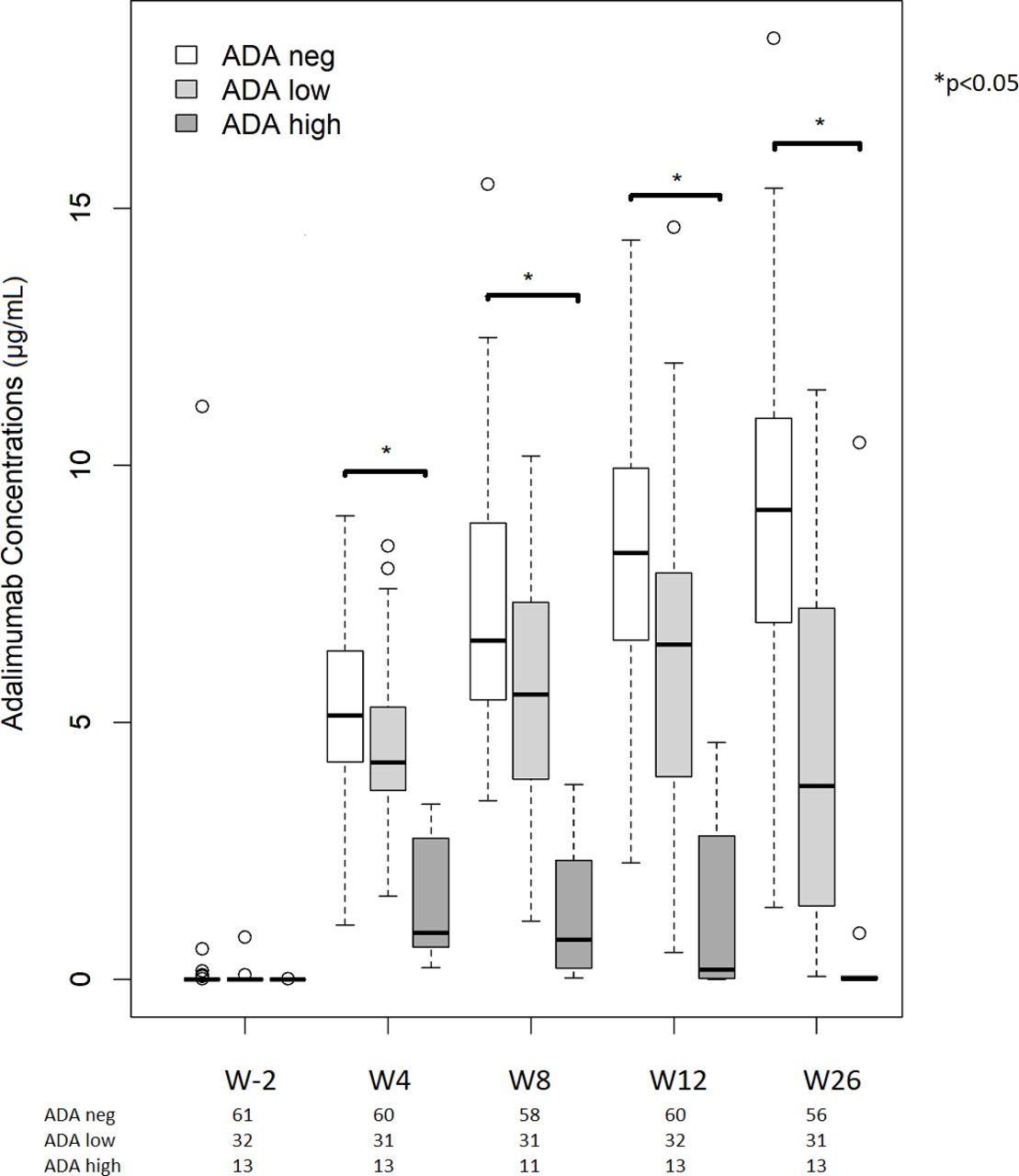
Samples were available at each visit as follows: W-2, n=106; W4, n=104; W8, n=100; W12, n=105; and W26, n=102. Adalimumab concentration was higher in the MTX+ than MTX− group after W-2 (figure 1). In linear mixed-effect models, MTX treatment was associated with higher adalimumab concentration at any time point (p<0.05). Median adalimumab concentration was lower for ADA-positive than ADA-negative patients at W26 (1.43 µg/mL (0.00–11.47) vs 8.66 µg/mL (0.05–18.31); p<0.05). In addition, adalimumab concentration was lower with ADA-high than ADA-low status throughout the study, the difference being apparent as early as W4 (figure 2).
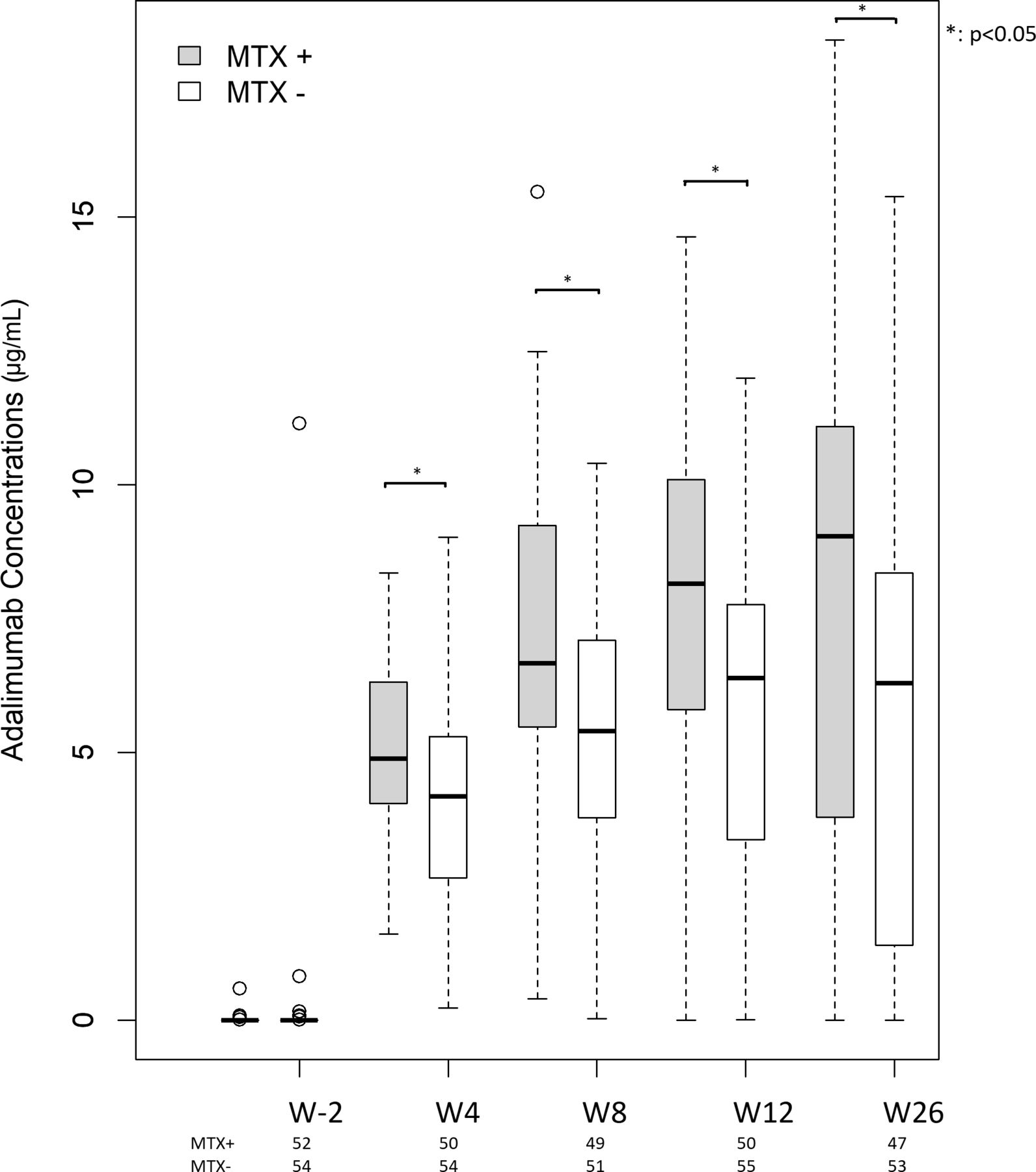
Adalimumab concentrations by treatment group. MTX+: methotrexate+adalimumab. MTX−: adalimumab alone. W-2=baseline, W4=4 weeks, W8=8 weeks, W12=12 weeks, W26=26 weeks. Four patients had a baseline adalimumab concentration above the lower limit of quantification because they previously received infliximab, which interfered with adalimumab detection. Horizontal lines are median, box edges are IQR and whiskers are range. The difference was statistically significant at W4, W8, W12 and W26.

Adalimumab concentrations by anti-drug antibody (ADA) status. MTX+: methotrexate+adalimumab. MTX−: adalimumab alone. W2=baseline, W4=4 weeks, W8=8 weeks, W12=12 weeks, W26=26 weeks. Horizontal lines are median, box edges are IQR and whiskers are range. The difference was statistically significant between ADA-high and ADA-low at W4, W8, W12 and W26.
Evolution of ASDAS and CRP level
In both groups, the ASDAS decreased at W4 and thereafter. At W26, the median ASDAS was 1.6 and was similar in the two treatment groups. The proportion of ASDAS responders at W26 was similar in the MTX− and MTX+groups (31/50 (62%), five missing data, and 25/46 (54%), six missing data; p=0.6). Likewise, the proportion of patients fulfilling ASDAS criteria for inactive disease was 40% (21/52, 3 missing data) and 37% (17/46, 6 missing data) in the MTX− and MTX+ groups, respectively (p=0.9). CRP level decreased during the study period, with no difference between the two groups (online supplementary figure 2).
Supplemental material
ASDAS was higher for ADA-positive than ADA-negative at W26 but not significantly (1.9 vs 1.5, p=0.06; data not shown). In addition, the clinical response was poorer for the ADA-high than ADA-low and ADA-negative patients (figure 3). In a linear mixed-effect model, ASDAS was higher for ADA-high than ADA-negative patients at the last visit (p=0.002).
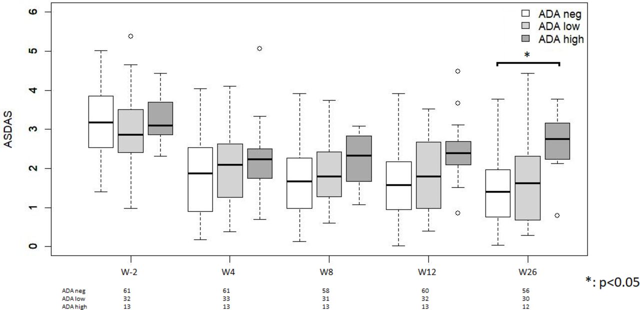
Ankylosing Spondylitis Disease Activity Score (ASDAS) by anti-drug activity (ADA) status. W0=0 week, W4=4 weeks, W8=8 weeks, W12=12 weeks, W26=26 weeks. The difference was only statistically significant between ADA-neg and ADA-high at W26.
Safety
The overall incidence of adverse events was similar in the two treatment groups (online supplementary table 1). Two serious adverse events were reported in each group. In the MTX− group, one patient had a paradoxical psoriasis pustulosa and one had drug-induced liver injury attributed to tuberculosis prophylaxis. In the MTX+ group, one patient presented pneumonia and then lower-limb ischaemia that resulted in hallux amputation. Further investigations led to the discovery of a foramen ovale with an atrial septal aneurysm. This patient was ADA-negative. The second patient was randomised in the MTX+ group and withdrew from the study because of nausea, diarrhoea and elevated liver enzyme activity, which was attributed to the combination of MTX and tuberculosis prophylaxis. The symptoms and blood test findings improved with treatment discontinuation and the patient was excluded from the statistical analysis.
Six patients (11%) in the MTX− group showed injection skin reactions as compared with one (2%) in the MTX+ group: four (57%) were ADA-positive as compared with 35/100 (35%) who did not present such reactions. In the four ADA-positive patients with skin reactions, three had ADA-low and one ADA-high status. The patient in the MTX+ group who presented with a skin reaction was ADA-positive and ADA-low.
Adalimumab long-term maintenance
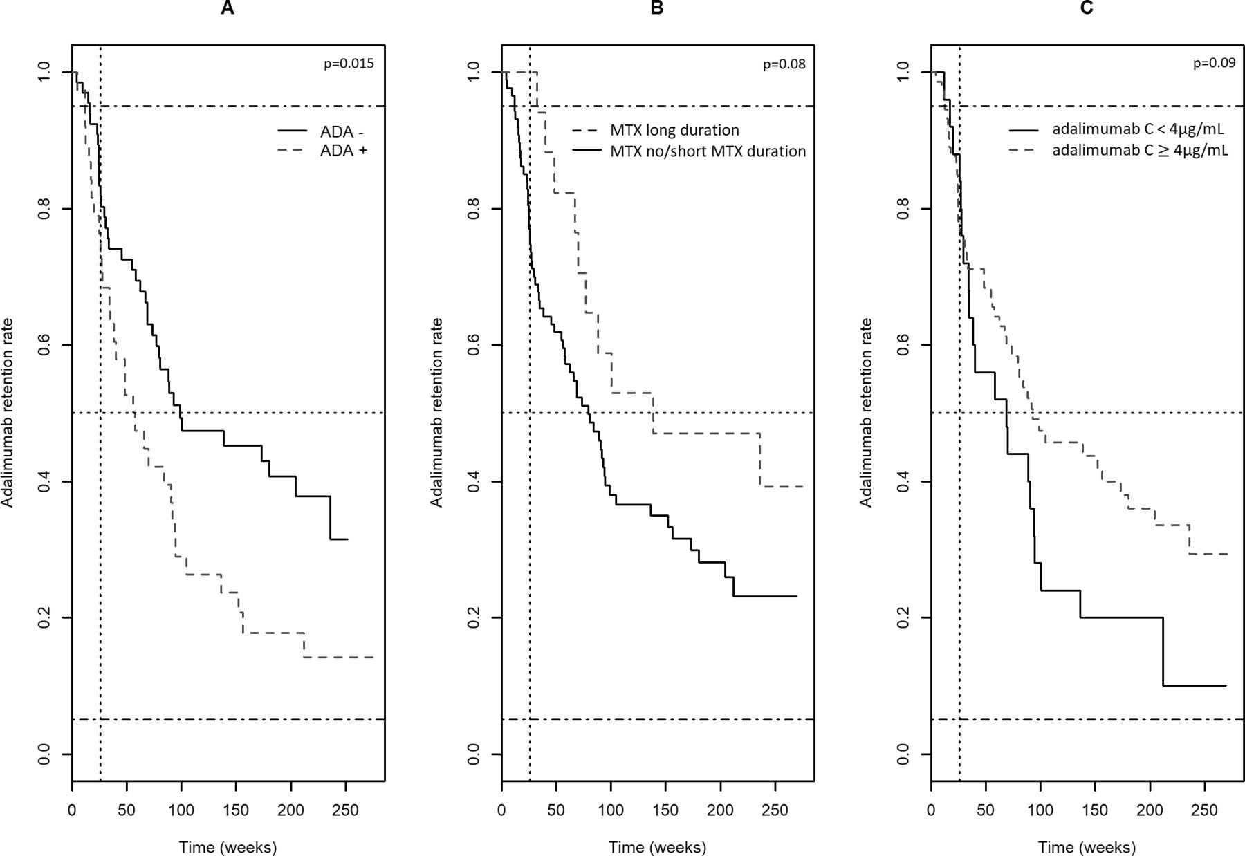
In the follow-up study, data were available for 104 patients: 50 in the MTX+ group and 54 in the MTX− group (online supplementary figure 1). The median follow-up was 210 weeks (25.3–274.7). The median survival of adalimumab was 88 weeks (4.3–236.0). Seventy patients discontinued adalimumab during the follow-up period. In the univariate analysis, ADA positivity at 6 months was the only covariate statistically associated with poor maintenance of adalimumab. The median survival of adalimumab in ADA-positive patients was 56.9 weeks (5.0–212.1), compared with 98.6 weeks (4.3–236.0) in ADA-negative patients (p=0.015) (figure 4A). There was no difference in the long-term adalimumab maintenance between MTX+ and MTX− groups (data not shown). Seventeen patients received MTX after the end of the study (with a median MTX duration of 79.1 (28–246.3) weeks), while 33 discontinued beforehand, mostly because of the non-approval of MTX in axial SpA, outside the clinical trial. There was a trend towards a longer adalimumab maintenance in patients with MTX long duration (>W26) as compared with those without MTX or short MTX duration (W26), the median survival of adalimumab being 138.6 weeks (32.4–236.0) and 79.6 weeks (4.3–212.1), respectively (p=0.08) (figure 4B). Maintenance of adalimumab in patients within the first quartile of adalimumab concentrations (<4 µg/mL) at W8 was poorer than in others (median survival of 68.6 weeks vs 92.7 weeks, respectively, p=0.09) (figure 4C). Adalimumab concentrations measured at W12 and 26 were not statistically associated with adalimumab long-term maintenance. In the Cox model multivariate analysis, including as covariables male versus female, absence versus presence of ADA at W26 and MTX co-treatment >W26 versus no MTX or short MTX duration (≤W26), the two variables statistically associated with the long-term maintenance of adalimumab were male sex and MTX >W26 (p=0.01 and p=0.04, respectively). The absence of ADA was not statistically associated with longer maintenance (p=0.07).

Adalimumab maintenance according to (A) anti-drug antibody (ADA)+ versus ADA− at week 26 (W26), (B) methotrexate (MTX) long duration, that is >W26, versus no MTX or MTX short duration, that is ≤W26 and (C) adalimumab concentrations<1st quartile at W8, that is <4 µg/mL, versus adalimumab concentration ≥1st quartile, that is ≥4 µg/mL.
Thus, MTX co-treatment duration >W26 or being a male halved the risk of discontinuing adalimumab, with a HR of 0.46 (0.22–0.95) and 0.51 (0.31–0.85), respectively. As opposed to this, being ADA positive at W26 did not significantly increase the odds of discontinuation of adalimumab (HR 1.57 (0.95–2.58)).
Discussion
In this prospective randomised trial, we demonstrate that MTX significantly reduced the immunogenicity and ameliorated adalimumab concentration, as a surrogate of drug exposure, in axial SpA patients. This result is of importance because immunogenicity to monoclonal antibodies is an unwanted outcome responsible for loss of response and treatment discontinuation in chronic inflammatory diseases.
The magnitude and persistence of an antibody response to a therapeutic protein is responsible for increased elimination and therefore decreased serum concentrations of the bioactive form of the drug.15 Adalimumab concentration was lower for patients with a high antibody response (ie, ADA level >100 AU/mL) than low or no antibody response, although not significantly. Hence, 10 mg MTX s.c. every week may reduce the risk of immunisation as a whole and prevent the risk of a high degree of immunisation, which has a negative impact in terms of pharmacokinetics and, in some cases, can trigger infusion reactions.16 However, the proposed scheme could not completely abrogate the immunogenicity of adalimumab in all MTX-treated patients because 25% were ADA-positive after W26, despite good adherence to MTX. The individual factors and mechanisms by which MTX reduces adalimumab immunogenicity deserve further studies.
MTX is not recommended for treating axial SpA. In an open-label study, 20 ankylosing spondylitis patients received high s.c. doses of MTX, and the effect did not differ from what was previously observed in the placebo group in a TNFi controlled trial.17 However, MTX is often used for SpA patients with peripheral signs, which provides some retrospective data concerning the effect of MTX on TNFi immunogenicity. In a retrospective study of infliximab in SpA patients, we found that those who received MTX concomitantly (n=25) were all ADA-negative, whereas 14/52 (27%) of those who did not receive MTX were ADA-positive.11 In their cohort, Plasencia et al found greater frequency of ADA to infliximab in patients who did not take MTX than in those with MTX combination therapy (20/58; 34.5% vs 4/36; 11.1%).12 Finally, Keepkens et al reported ADA to adalimumab in 27% of ankylosing spondylitis patients at week 24 and in none of the five patients who concomitantly used MTX.5 The present randomised trial demonstrates that MTX reduced adalimumab immunogenicity in axial SpA and suggests a potential benefit of this combination.
The choice of the MTX dose, initiation time and route of administration was a compromise between the expected immunological effect and acceptable tolerance. Krieckaert et al reported that concomitant MTX at low dosage (5–10 mg/week), intermediate dosage (12.5–20 mg/week) or high dosage (≥22.5 mg/week) dose-dependently decreased the percentage ADA detection in rheumatoid arthritis patients: at week 28, the proportion of ADA-positive patients without MTX was ~45% versus ~10% for patients with moderate-dose MTX.10 These data were later confirmed in the CONCERTO trial, the percentage of ADA-positive patients being 6% in both the 10 and 20 mg MTX dose groups, as compared with the 2.5 mg (21%) and 5 mg (13%) MTX dose groups.18
MTX bioavailability of oral and s.c. administration has been studied in rheumatoid arthritis patients receiving ≥25 mg/week, demonstrating a higher area under the concentration curve (AUC) with s.c. administration and a positive dose–AUC relation as compared with oral administration.19 This dose-dependent linear increase in drug exposure was later confirmed by Schiff et al, who concluded to no pharmacokinetic advantage in increasing the oral dose of MTX above 15 mg/week,20 which is the evidence-based recommended dosage for rheumatoid arthritis.21 Hence, based on the reduced immunogenicity observed in rheumatoid arthritis patients,10 we chose the 10 mg/week s.c. regimen in this study. According to the method recently established by Schiff et al, this dosage corresponds to ~12.5 mg/week oral dosage, a regimen that probably would have yielded similar results, with a much lower cost than the s.c. route.22 Most importantly, the parenteral route is known to improve tolerance and therefore, adherence to MTX, which may have by itself contributed to the reduced immunogenicity.23 The rather low 10 mg/week dose regimen may however account for the residual immunogenicity observed in 25% of the MTX+ group, rising the hypothesis that some patients may have deserved a higher or weight-adjusted dose. Finally, MTX was initiated 2 weeks before adalimumab initiation to maximise its effect on reducing the immune response. The CONCERTO trial demonstrated recently that starting both MTX and adalimumab simultaneously was also able to reduce ADA development.18
One important finding is the enhanced adalimumab trough concentration, a surrogate of drug exposure, in the combination group as compared with adalimumab monotherapy. This finding was reported in rheumatoid arthritis,24 and might be attributed to two mechanisms. First, MTX may have a direct immunosuppressive effect on the humoral response to adalimumab, thus decreasing the magnitude and length of ADA production.25 Second, MTX co-medication, which is associated with a 30% decrease in clearance of infliximab in rheumatoid arthritis,26 may have resulted in an early high serum concentration of adalimumab in our study, thereby leading to lower immunogenicity in the MTX+ than MTX− group.27 In an animal model, some authors have recently observed an increased FcRn expression in tissues, along with a decrease of adalimumab clearance in MTX-treated rats, as compared with animals not receiving MTX.28 Thus, MTX may have resulted in an increased expression of FcRn expression, which contributed to increased adalimumab concentration, in our study. Further pharmacokinetics and pharmacokinetic-pharmacodynamics analyses are required to explore these hypotheses. The precise mechanisms of action of MTX and its metabolites on ADA development deserve more comprehensive further analyses. In terms of clinical practice, the dose tapering of adalimumab could be facilitated in those patients with high trough concentration as was recently observed in rheumatoid arthritis,29 which supports the potential interest of combining MTX to adalimumab in axial SpA. The existing evidence in rheumatoid arthritis is however still lacking in axial SpA, in this respect.
The two treatment groups did not differ in clinical response or CRP level at the end of the prospective study. However, ADA-positive patients showed low adalimumab concentration and poor response, particularly those with high immunisation level. The quantification of the effect of MTX on clinical outcomes such as ASDAS, should be assessed further than 26 weeks, in future studies. In our follow-up study, we observed a longer maintenance of adalimumab in male, which aligns with the data from the Danish nationwide DANBIO registry.30 With regard to MTX co-treatment, our results are in accordance with the effect of prolonged MTX on adalimumab maintenance reported by Lie et al.31 In our study MTX was maintained in some patients after the end of the 26-week study period, which enabled us to compare the maintenance of adalimumab in these patients with those who discontinued MTX beforehand. Although the difference was not statistically significant, the data argue for a continuous effect of MTX on adalimumab maintenance. Finally, we observed a trend towards a poorer maintenance of adalimumab in patients whose concentration at W8 was less than 4 µg/mL, which is in agreement with our previous work in SpA with infliximab.32 The patients were not randomised to maintain or discontinue MTX, which could have somewhat biased our results. The number of covariates in the Cox model was limited to the most documented factors from the literature. Several other factors could have also influenced the treatment maintenance but, owing the limited number of patients, only sex, MTX co-medication and ADA were studied in this work.
The association of ADA positivity and risk of reactions to adalimumab injection was previously reported by Pascual-Salcedo et al, particularly in patients with high ADA titres.16 In our study, the incidence of reactions was increased with ADA positivity. However, a large number of these cases could not be explained by immunogenicity, as assessed by the antigen-binding test.
Conclusion
Starting MTX 10 mg/week s.c. 2 weeks before adalimumab may reduce the immunogenicity and ameliorate the pharmacokinetics of adalimumab in axial SpA patients. Although combining MTX and adalimumab does not improve the clinical response after W26, the reduced ADA positivity and infusion reaction, along with the increase in adalimumab serum concentration, should benefit axial SpA patients. The prolonged co-treatment of MTX with adalimumab may therefore improve the therapeutic maintenance of adalimumab. Further studies are needed before the present findings can be extrapolated to other TNFi.
Supplemental material
Acknowledgments
The authors thank Bruno Giraudeau for methodological advice in the study design; Yoann Desvignes for technical support with the study protocol; Elody Marnat for data management; Anne-Claire Duveau and Caroline Brochon for technical assistance in measuring adalimumab concentrations, and Kim van Houten and Asma Kalei (Sanquin Diagnostic Services, Amsterdam, Netherlands) for measuring anti-drug antibodies to adalimumab. We thank Sophie Guyétant (clinical research manager), Coraline Gadras (Centre de ressource Biologique-Biobank), Annie-Pierre Jonville-Bera (Pharmacovigilance), Emmanuelle Mercier, Anne Cécile Henriet and Maud François (committee for evaluation of safety). Measurement of adalimumab serum concentrations was performed within the 'Centre pilote de suivi biologique des anticorps thérapeutiques' (CePiBAc) – Pilot centre for therapeutic antibodies monitoring platform of Tours University Hospital, which was cofinanced by the European Union. Europe is committed to the region Centre with the European Regional Development Fund (ERDF). This work was partly supported by the French Higher Education and Research Ministry under the program Investissements d’avenir (grant agreement: LabEx MAbImprove ANR-10-LABX-53-01). This work was a collaborative venture by the VICTOR HUGO network (Hôpitaux Universitaires du Grand Ouest—Western France University Hospitals; http://www.srouest.fr), dedicated to innovative research on rheumatic diseases. DM, PG, GP, CD, DT, TR and AdV participated in the Consortium ‘Monitoring of monoclonal Antibodies Group in Europe’ (MAGE) for inflammatory diseases. LE STUDIUM Loire Valley Institute supports the MAGE Consortium for Advanced Studies (http://www.lestudium-ias.com/). We thank Laura Smales (BioMedEditing) for improving the English language and editing the manuscript. We are in debt to the people who committed themselves to the recruitment, clinical evaluation, blood sampling, drug dispensation and data collection in each centre. Brest: Thierry Marhadour (co-investigator), Sandrine Jousse-Joulin (co-investigator), Alain Saraux (co-investigator), Divi Cornec (co-investigator), Sophie Varache (co-investigator), Christine Guimard (pharmacist), Claire Poullaouec (pharmacist), Emmanuelle Eleouet (pharmacist), Nicole Thiébaut (pharmacist), Elsa Menanteau (clinical study technician), Liana Le Roux (clinical study and lab technician), Clarysse Adrien (clinical study technician), Nathalie Bihannic (clinical study technician), Stéphanie Paul (nurse), Marie-Louise Guenegues (nurse), Marie Jezequel (clinical study technician), Alice Rameur (Nurse). Blois: Marie-Pierre Kuzzay (pharmacist), Philippe Breton, (pharmacist). Le Mans: Guillaume Direz (co-investigator), Frédéric Medina (co-investigator), Amélie Denis (co-investigator), Vincent André (co-investigator), Anne-Marie Caminondo (pharmacist), Anne-Marie Vidal (pharmacist), Delphine Bollé (pharmacist) Lodna Jemour (pharmacist), Eglantine Rouanet (clinical research assistant), Angélique Royer (lab technician), Fabienne Daneuil Potier (nurse), Isabelle Polome (nurse). La Roche-sur-Yon: Céline Cozic (co-investigateur), Céline Chantreau (clinical research assistant), Nadine Rabillé (clinical study technician), Yannick Poirier (pharmacist), Marie-Line Martin (pharmacy), Stéphane Varin (co-investigator), Gilles Tanguy (co-investigator), Michel Caulier (co-investigator), Isabelle Lepage (co-investigator), Laurence Bessonnet (study nurse), Emeline Gaigneux (co-investigator). Nantes: Yves Maugars (co-investigator), Jean-Marie Berthelot (co-investigator), Laurent Flet (pharmacist), Julie Moynard (pharmacist), Karen Batard (clinical study technician), Peggy Ageneau (clinical study technician). Orléans: Carine Salliot (co-investigator), Sylvie Loiseau-Peres (co-investigator); Béatrice Gareau (clinical research assistant), Corinne Ruet (clinical research nurse), Farida Khacef (clinical study technician), Sophie Tollec (pharmacist). Poitiers: Christine Bécuet (pharmacist), Katia Gourou (clinical research assistant), Isabelle Azais (co-investigator), Julien Girodon (co-investigator), Géraldine Durand (co-investigator), Céline Thomas (clinical study technician). Rennes: Anne-Laure Deniau (pharmacist), Pauline Lepot (pharmacist), Anne Mucheron (pharmacist), Christine Bloino-Ayoul (research nurse), Guillaume Coiffier (co-investigator), Stéphan Pollet (co-investigator), Thomas Bourrée (co-investigator), Christine Mot (Research nurse). Saint-Nazaire: Olivier Lemenand (biologist), Laurent Ott (administrator), Alix Phelizot (Clinical research assistant), Aurore Deininger (pharmacy assistant), Oriane Mérot (co-investigator). Saint-Brieuc: Marie-Cécile Hervé (clinical research assistant). Tours: Virginie Martaillé (co-investigator), Valérie Védère (co-investigator), Saloua Mammou (co-investigator), Isabelle Griffoul (co-investigator), Dominique Soutif (co-investigator), Julien Mélet (co-investigator), Hélène Bourgoin (pharmacist), Thibault Page (pharmacist), Françoise Girault (pharmacist assistant), Stéphanie Bonte (clinical research assistant), Nicolas Charron (clinical study technician), Lysiane Brick (clinical research nurse). We thank John Isaacs (Newcastle, UK) and Maxime Dougados (Paris, France) for reading the manuscript and offering their advice.
References
Footnotes
Contributors EDu contributed to the study conception and design, performed the statistical analysis, participated in the patients’ selection, clinical assessment and data collection, interpreted the results and drafted the manuscript. DM was responsible for coordination, conception and design of the study, participated in the patients’ selection, clinical assessment and data collection, interpreted the results and drafted the manuscript. MS contributed to the posthoc survival analysis, statistical analysis and drafted the manuscript. PG contributed to the study conception and design, participated in the patients’ selection, clinical assessment and data collection and participated in interpretation of results. eDe, FLG, LA, AP, EL, AM, GC, TA, VD-P, EG, and BLG improved the study design, participated in the patients’ selection, clinical assessment, data collection and interpretation of results. TR and AdV were responsible for antibodies towards adalimumab detection and interpreted the results. CD was responsible for serum sample management, material transfer and interpreted the results. DT was responsible for adalimumab measurement and participated in interpretation of results. EP was responsible for c-reactive protein measurement participated in interpretation of results. GP and HW participated in interpretation of results and critically reviewed the manuscript. All authors revised and approved the final manuscript.
Funding This work was promoted by the Regional University Hospital Centre of Tours and supported by grants from the French Ministry for Health and Sport within the framework of the Programme Hospitalier de Recherche Clinique 2012. The authors thank Hervé Brunet, Véronique Dubreuil-Chambardel and Patrick Thiennot from the Lions Club, Tours Val de France for their support and funding, and Nordic Pharma, which provided the methotrexate syringes.
Competing interests EDu was invited to attend international congresses by Roche and UCB; she has acted as a consultant and given lectures on behalf of her institution for BMS and Abbvie. eDe participated on behalf of her institution in clinical trials sponsored by Abbvie, Roche, BMS, Novartis, Pfizer, UCB and Sanofi; she has given lectures for Abbvie, BMS, Janssen, Pfizer, UCB, Novartis; she has acted as a consultant for BMS and UCB, Novartis; she has been invited to attend international congresses by MSD, Roche, BMS Abbvie and Novartis. FLG has been invited to attend an international congress by Abbvie, Pfizer. LA was invited to attend international congress by Abbvie, Novartis, Pfizer and UCB. EL has received speaker and consultant fees from Amgen, Expanscience, Lilly, and MSD; and research grants from Abbvie, Amgen, Lilly, MSD and UCB. GC was invited to attend international congress by Abbvie. VD-P has received speaker and consultant fees from MSD, BMS, UCB, Roche; and research grants from Roche-Chugai. EG has participated on behalf of his institution in clinical trials sponsored by Roche, Lilly, Novartis, Amgen, and BMS; she has acted as a consultant and given lectures for Abbvie, BMS, MSD, Pfizer, Roche, UCB, Novartis; she has been invited to attend international congresses by MSD, Roche, Novartis and BMS. BLG has participated on behalf of his institution in clinical trials sponsored by Roche, Lilly, Novartis, Pfizer, UCB and MSD; he has acted as a consultant and given lectures for Abbvie, BMS, Janssen, MSD, Pfizer, Sanofi-Genzyme, UCB, Novartis; he has been invited to attend international congresses by MSD, Roche, Abbvie, Sanofi and Pfizer. EP was invited to attend an international congress by Buhlmann. GP reports grants received by his research team from Novartis, Roche Pharma, Sanofi-Genzyme, Chugai, Pfizer and Shire, outside of the submitted work. DT has acted as a consultant and given lectures for Sanofi, Amgen, PG participated on behalf of his institution in clinical trials sponsored by Abbvie, Roche, BMS, Boehringer, Lilly, Novartis, Pfizer, UCB, Janssen and MSD; he has acted as a consultant and given lectures for Abbvie, Biogaran, BMS, Hospira, Janssen, MSD, Pfizer, Sanofi-Genzyme, UCB; he has been invited to attend international congresses by MSD, Roche, BMS and Abbvie. DM has acted as a consultant and given lectures on behalf of his institution for Pfizer and Novartis; he has been invited to attend an international congress by Janssen-Cilag. His institution received grants for research from the non-governmental organisation Lions Club Tours Val de France. TR, MS, AP, CD, AdV, TA, AM and HW declared that they have no disclosure with the manuscript.
Patient consent for publication Not required.
Ethics approval This study was approved by the ethics committee of Tours University Hospital and was conducted in accordance with the Declaration of Helsinki. It was registered at ClinicalTrials.gov (NCT01895764).
Provenance and peer review Not commissioned; externally peer reviewed.
Data availability statement Data may be obtained from a third party and are not publicly available.