Article Text

Abstract
Objectives To characterise peripheral musculoskeletal involvement in patients with spondyloarthritis (SpA) including psoriatic arthritis (PsA), across the world.
Methods Cross-sectional study with 24 participating countries. Patients with a diagnosis of axial SpA (axSpA), peripheral SpA (pSpA) or PsA according to their rheumatologist were included. The investigators were asked which diagnosis out of a list of six (axSpA, PsA, pSpA, inflammatory bowel disease-associated SpA, reactive arthritis or juvenile SpA (Juv-SpA)) fitted the patient best. Peripheral manifestations (ie, peripheral joint disease, enthesitis, dactylitis and root joint disease), their localisation and treatments were evaluated.
Results A total of 4465 patients were included (61% men, mean age 44.5 years) from four geographic areas: Latin America (n=538), Europe plus North America (n=1677), Asia (n=975) and the Middle East plus North Africa (n=1275). Of those, 78% had ever suffered from at least one peripheral musculoskeletal manifestation; 57% had peripheral joint disease, 44% had enthesitis and 15% had dactylitis. Latin American had far more often peripheral joint disease (80%) than patients from other areas. Patients with PsA had predominantly upper limb and small joint involvement (52%).
Hip and shoulder involvement was found in 34% of patients. The prevalence of enthesitis ranged between 41% in patients with axSpA and 65% in patients with Juv-SpA. Dactylitis was most frequent among patients with PsA (37%).
Conclusion These results suggest that all peripheral features can be found in all subtypes of SpA, and that differences are quantitative rather than qualitative. In a high proportion of patients, axial and peripheral manifestations coincided. These findings reconfirm SpA clinical subtypes are descendants of the same underlying disease, called SpA.
- spondylitis
- ankylosing
- arthritis
- psoriatic
- arthritis
- juvenile
This is an open access article distributed in accordance with the Creative Commons Attribution Non Commercial (CC BY-NC 4.0) license, which permits others to distribute, remix, adapt, build upon this work non-commercially, and license their derivative works on different terms, provided the original work is properly cited, appropriate credit is given, any changes made indicated, and the use is non-commercial. See: http://creativecommons.org/licenses/by-nc/4.0/.
Statistics from Altmetric.com
Key messages
What is already known about this subject?
Peripheral musculoskeletal manifestations are a hallmark of psoriatic arthritis (PsA) and occur also in spondyloarthritis (SpA), but their distribution and worldwide prevalence have sparsely been studied in SpA and specifically in axial SpA (axSpA).
What does this study add?
This study demonstrates that all types of peripheral musculoskeletal manifestations can be found in all subtypes of SpA.
This study allowed to directly compare the worldwide SpA (including PsA) population across the different geographic areas, and demonstrated a high inter-region variability in the prevalence of these manifestations.
There is a large overlap of peripheral manifestations across the different SpA subtypes, suggesting a high prevalence of peripheral features in axSpA and quite similar prevalences in peripheral SpA and PsA.
How might this impact on clinical practice or future developments?
This study suggests that SpA constitutes one entity with different phenotypic presentations (including PsA).
This study confirms that peripheral musculoskeletal manifestations should be studied in the entire group of SpA rather than in its subgroups alone.
Introduction
Spondyloarthritis (SpA) mainly affects the axial skeleton and sacroiliac joints but may affect peripheral structures too.1 Peripheral involvement has always been important in the nomenclature of the diverse forms of SpA. The traditional classification includes several subtypes, such as ankylosing spondylitis (AS), psoriatic arthritis (PsA), inflammatory bowel disease (IBD)-associated SpA, reactive arthritis (ReA) and juvenile SpA (Juv-SpA), depending on the presence of peripheral and/or extramusculoskeletal manifestations.1 In 2009, the Assessment of Spondyloarthritis international Society (ASAS) has introduced the concept of axial (axSpA) and peripheral SpA (pSpA) and developed new classification-criteria making this distinction.2 In parallel, and independently, the Group for Research and Assessment of Psoriasis and Psoriatic Arthritis (GRAPPA) has proposed specific criteria for the classification of PsA.3 Guided by the reality of drug development in the field, the US Food and Drug Administration and the European Medicine Agency have proposed specific guidance for drug development limited to three (four) diseases (axSpA (radiographic and non-radiographic (AS)), pSpA and PsA), which to some extent ignores the reality of SpA as an umbrella entity. This pleiotropy has created dilemmas in the field, since patients with peripheral manifestations, that can occur across the entire spectrum of patients with SpA, may easily meet the criteria for more than one disease at the same time, or alternatively may fit distinct criteria over time. Similarly, patients with a SpA-associated diagnosis characterised by peripheral involvement may also present with axial symptoms. Finally, it is suggested that distributions of peripheral manifestations of SpA may differ across geographical areas. It is well known, for example, that the prevalence of peripheral arthritis in Latin American patients with SpA is higher than in Europe4 5 and that peripheral involvement of SpA in Asia is rather low.6
The study of phenotypical distributions has classically evolved along the axes of scientific and professional organisations (eg, GRAPPA vs ASAS), which focused on either peripheral or axial involvement. In addition, ASAS classically had a European dominance, while GRAPPA had a solid North American basis. Moreover, in the majority of previous SpA cohorts, these manifestations (either axial or peripheral) were used as entry criterion, limiting the evaluation of the whole SpA spectrum. These distinctions imply and explain why a thorough comparison of peripheral manifestations in the SpA spectrum, with involvement of countries around the world, has never been conducted so far. This has prompted us to design the ASAS-PerSpA study, which aimed at comparing the prevalence, characteristics and treatments of peripheral musculoskeletal manifestations in patients with all possible subtypes of SpA across all regions of the world. The investigators were also interested to see how often peripheral and axial manifestations in patients with a SpA diagnosis coincide, and if there are global differences.
Methods
Study design
PerSpA was a multinational observational, cross-sectional study with 24 participating countries worldwide.
Patient recruitment
For this study, the scientific committee appointed one national principal investigator (an ASAS member) for each participating country. Several countries per continent were selected and the national principal investigators invited rheumatologists from their countries to participate. Consecutive adult patients (ie, at least 18 years old) with a diagnosis of axSpA, pSpA or PsA, who were able to understand and complete questionnaires, were included from July 2018 to February 2020.
Data collection
A specific case report form was used to collect four different categories of data:
Demographics: country, age, sex, body mass index, smoking, alcohol intake and the highest level of education completed.
Disease characteristics: the investigators were asked to name the diagnosis that in their opinion best described the disease of the patient irrespective of the fulfilment of any classification criteria. They could choose from the following list: axSpA, PsA, pSpA, IBD-related SpA, ReA, Juv-SpA or they could name another disease.
In addition, information about HLA-B27 status, first-degree or second-degree relatives (with AS, psoriasis, uveitis, ReA or IBD), axial involvement (defined with the question “Do you consider that this patient has ever suffered from axial involvement of SpA?”), information concerning the presence of sacroiliitis on radiographs, uveitis, psoriasis confirmed by a dermatologist, IBD confirmed by endoscopy and treatment (non-steroidal anti-inflammatory drugs (NSAIDs), glucocorticoids, conventional synthetic (cs) and biological (b) disease-modifying antirheumatic drugs (DMARDs)) were collected.
Peripheral musculoskeletal manifestations: included (a) peripheral joint disease (excluding root joints) in the past, the presence of objective signs of synovitis (ie, physical examination by a rheumatologist or confirmed by ultrasonography), a monoarticular, oligoarticular or polyarticular pattern, localisation (predominantly in the lower limbs/large joints) and natural history (transient, continuous, intermittent or progressive); (b) ‘root-joint’ (ie, hip and shoulder) involvement in the past according to the rheumatologist, (c) midfoot arthritis (tarsitis) in the past as well as confirmed by specific investigations; (d) enthesitis in the past confirmed and non-confirmed by specific tests (ie, sonography, radiographs, MRI or bone scintigraphy), localisation and natural history (single episode, intermittent, continuous or progressive) and (e) the occurrence of dactylitis in the past and localisation (fingers or toes) were collected. Moreover, the presence of current peripheral musculoskeletal manifestations at the moment of the study visit was evaluated based on physical examination. Investigators were also asked about specific treatments (NSAIDs, oral and intra-articular glucocorticoids, csDMARDs and bDMARDs) prescribed for each of these peripheral musculoskeletal manifestations using the questions ‘Did this manifestation require a specific treatment?’ and ‘if yes, which one?’
Clinimetric information: current disease activity at the moment of the study visit was measured by the Bath Ankylosing Spondylitis Disease Activity Index7 and the Ankylosing Spondylitis Disease Activity Score-C reactive protein (ASDAS-CRP).8 Moreover, the Ritchie Articular Index,9 66 Swollen Joints Index,10 Mander enthesitis index (MEI),11 Leeds Enthesitis Index12 and the Spondyloarthritis Research Consortium of Canada enthesitis score13 were assessed. The Bath Ankylosing Spondylitis Functional Index and the ASAS Health Index were used to evaluate function and health, respectively.14 15 Finally, the presence of secondary fibromyalgia according to the rheumatologist’s opinion was collected and the self-reported Fibromyalgia Rapid Screening Tool (FiRST) was completed.16
All information was obtained by a study investigator or research nurse during a face-to-face interview at one single study visit, which included a review of the medical record. A centralised electronic case report form was used to enter the data.
Statistical analysis
Patients were stratified in four geographic areas: Latin America (Argentina, Chile, Colombia and Mexico), Europe and North America (Canada, France, Germany, Hungary, Italy, Portugal, Romania, Spain, the Netherlands, the UK and the USA), Asia (China, India, Japan, South Korea and Taiwan) and the Middle East and North Africa (Egypt, Lebanon, Morocco and Turkey). North America and Europe were grouped together because the numbers of patients from the USA and Canada were low.
Patients were also grouped according to the best-fit diagnosis (axSpA, PsA, pSpA, IBD-SpA, ReA, Juv-SpA or alternatives). The analysis was purely descriptive. The prevalence of each peripheral musculoskeletal manifestation was estimated and information about such manifestations (locations, natural history and specific treatments) was examined with regard to the geographic area and to the diagnosis.
Results
Demographics and disease characteristics
Investigators from 24 countries recruited a total of 4538 consecutive patients fulfilling the inclusion criteria. Seventy-three patients were excluded because of missing data, and 4465 patients were included in the analysis. The number of patients included per country is summarised in online supplemental table S1. Latin America (12%), Europe and North America (38%), Asia (22%) and the Middle East plus North Africa (29%) were properly represented. North America and Europe were grouped together because the enrolled patients in the USA and Canada were low (only 110 patients). The best-fit diagnoses (in decreasing order) was axSpA (61%), followed by PsA (23%), pSpA (9.7%), SpA-IBD (2.5%), ReA (1.3%), other SpA (1.3%) and Juv-SpA (1.2%). Demographics, clinical characteristics, disease activity and disease burden with regard to the diagnosis are shown in table 1. The prevalence of the HLA-B27 antigen ranged between 18.2% in patients with PsA and 79% in patients with axSpA. Uveitis was found less frequently in patients with PsA (2.6%) and more frequently in patients with axSpA (22%). Patients with PsA had the highest prevalence of psoriasis confirmed by a dermatologist (87%) while patients with ReA and IBD-SpA had the lowest prevalence (3%). Remarkably, 55% of patients with pSpA and 36% of patients with PsA had axial involvement according to the rheumatologist, and 52% of patients with axSpA had ever used csDMARDs. At the moment of the study visit, the prevalence of patients with low disease activity (ASDAS-CRP <2.1) ranged between 36% in pSpA and 41% in axSpA. Online supplemental table S2 shows the same information with regard to the geographic area. HLA-B27 was more frequent among Asian patients (80%) and less frequent among patients from Middle East and North Africa (54%). Asian patients also had the highest prevalence of axial involvement according to the rheumatologist (82%), while only 66% of Latin American patients had axial involvement. Finally, IBD was more prevalent among Middle East and North African patients (8%) and less prevalent among Asian patients (3%).
Supplemental material
Sociodemographics and clinical characteristics, disease activity and disease burden with regard to the diagnosis
Peripheral musculoskeletal manifestations
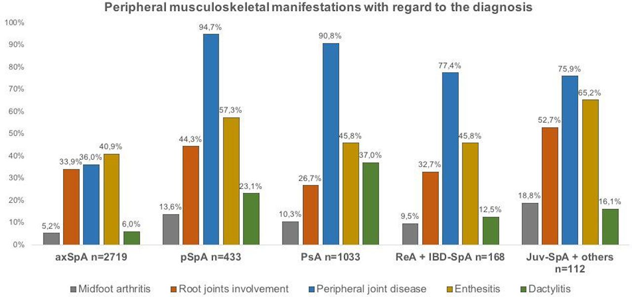
Of all patients, 78% had suffered at least once from a peripheral musculoskeletal manifestation (either peripheral joint disease, root joint involvement, tarsitis, enthesitis or dactylitis). The lowest prevalence was found in axSpA (66%), the highest expectedly in pSpA (99%). The prevalence of musculoskeletal manifestations was plotted against geographic area and diagnosis in figures 1 and 2, respectively. At the moment of the study visit, 32% of patients had at least one current peripheral musculoskeletal manifestation on physical examination (23% of patients with axSpA and 49% of patients with pSpA).

Prevalence of peripheral musculoskeletal manifestations in the past with regard to the geographic area.

Prevalence of peripheral musculoskeletal manifestations in the past with regard to the diagnosis. axSpA, axial spondyloarthritis; IBD-SpA, inflammatory bowel disease-associated spondyloarthritis; Juv-SpA, juvenile spondyloarthritis; PsA, psoriatic arthritis; pSpA, peripheral spondyloarthritis; ReA, reactive arthritis.
Peripheral joint disease (excluding root joints)
Peripheral joint disease (excluding root joints) was the most frequent peripheral musculoskeletal manifestation in the whole population with a prevalence of 57% (51% had objective signs of synovitis). The geographical distribution of peripheral joint disease differed importantly: patients from Latin American countries showed the highest prevalence (80%). According to the diagnosis, the lowest prevalence of peripheral joint disease was found in patients with axSpA (36%), while the highest frequency was found in patients with pSpA and PsA (95% and 91%, respectively).
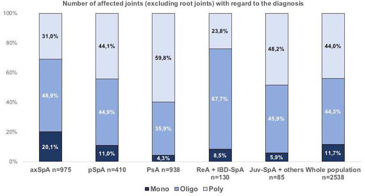
The number of affected joints with regard to diagnosis is illustrated in figure 3. Among the whole population with peripheral joint disease, 12%, 44% and 44% of patients showed monoarticular, oligoarticular and polyarticular involvement, respectively. But patients with a diagnosis of PsA had predominantly polyarticular involvement (60%), while patients with a diagnosis of ReA or IBD-SpA had predominantly oligoarticular disease (68%). Monoarticular involvement was rare in all groups.

Number of affected joints in the past (excluding root joints) with regard to the diagnosis*. *Among patients with peripheral joint disease (excluding root joints) and available data concerning the number of affected joints (n=2538). axSpA, axial spondyloarthritis; IBD-SpA, inflammatory bowel disease-associated spondyloarthritis; Juv-SpA, juvenile spondyloarthritis; PsA, psoriatic arthritis; pSpA, peripheral spondyloarthritis; ReA, reactive arthritis.
Of all patients with peripheral joint disease, 39% had predominantly lower limb and large joint involvement (figure 4); 31% had only peripheral joint disease of small joints of upper limbs (hands). Interestingly, the prevalence of patients with predominantly lower limb and large joint involvement was similar in patients with pSpA (51%) and axSpA (49%). Of the patients with a diagnosis of PsA, 52% had predominantly upper limb and small joint involvement.

Location of peripheral articular involvement in the past (excluding root joints) with regard to the diagnosis*. *Among patients with peripheral joint disease (excluding root joints) and available data concerning the location of affected joints (n=2501). axSpA, axial spondyloarthritis; IBD-SpA, inflammatory bowel disease-associated spondyloarthritis; Juv-SpA, juvenile spondyloarthritis; PsA, psoriatic arthritis; pSpA, peripheral spondyloarthritis; ReA, reactive arthritis.
Despite the use of bDMARDs at the moment of the study visit was more frequent among patients with axSpA than in pSpA (51.6% vs 47.5%, respectively), patients with pSpA showed the highest prevalence of at least one swollen joint on physical examination (42%) and patients with axSpA showed the lowest prevalence (10%) (table 1).
csDMARDs and bDMARDs specifically for peripheral joint disease were used in 77% and 42% of patients, respectively, while systemic glucocorticoids and local injections were used in 43% and 31%, respectively (table 2).
Specific treatments with regard to each peripheral musculoskeletal manifestation*
Root joint involvement
Root joint involvement (ie, hip or shoulder) occurred among all subtypes but ranged between 27% in patients with PsA and 53% in patients with Juv-SpA. Asian patients had most root joint involvement (55%) in comparison with the other regions. Among patients with root joint involvement and available data concerning the location (figure 5), hip involvement alone was found in 57%, being most frequent in axSpA (65%), whereas shoulder involvement alone was found in 21%, being more prevalent in patients with PsA (43%). The highest prevalence of hip and shoulder involvement occurring in the same individual was found in patients with pSpA (32%). Among patients with root joint involvement, 30% initiated bDMARDs specifically for this symptom, while 9% required total articular replacement (table 2).

Location of root joint involvement in the past with regard to the diagnosis*. *Among patients with root joint involvement and available data concerning the location (n=1372). axSpA, axial spondyloarthritis; IBD-SpA, inflammatory bowel disease-associated spondyloarthritis; Juv-SpA, juvenile spondyloarthritis; PsA, psoriatic arthritis; pSpA, peripheral spondyloarthritis; ReA, reactive arthritis.
Midfoot arthritis (tarsitis)
A total of 344 patients in the overall population had ever suffered from midfoot arthritis (tarsitis), representing 7.7% (2.2% confirmed by specific investigations). The prevalence of tarsitis ranged between 5.2% in patients with axSpA and 19% in patients with Juv-SpA. With regard to the region, this frequency ranged between 3.5% in Middle East and North African patients and 24% in Latin American patients.
Enthesitis
Of all patients, 44% had ever suffered from any enthesitis (17% had imaging-confirmed enthesitis). Enthesitis was more prevalent in Latin America (61%) than in other countries (approximately 40%). Enthesitis was more prevalent in patients with Juv-SpA (65%) than in other patients (approximately 45%).
The heel (either the insertion of the Achilles tendon or the plantar fascia) was by far the most frequent first location for enthesitis (69%). The course of enthesitis (online supplemental figure S1) was intermittent (55%), continuous (21%), monophasic (19%) or progressive (12%). The mean number of locations was 4.3 (4.6), and slightly higher in patients with a diagnosis of PsA (4.8 (5.3)) (online supplemental figures S2 and S3).
At the moment of the study visit, 18% of patients had enthesitis according to the MEI on physical examination (ie, at least one enthesis with a score >1), less often in patients with axSpA (17%) and more often in patients with ReA and IBD-SpA (26%) (table 1).
The specific treatment used for enthesitis is described in table 2. Remarkably, only 8.1% of patients received local injections with glucocorticoids, and 34% received csDMARDs specifically for this symptom.
Dactylitis
Of all patients, 15% had ever dactylitis. Expectedly, the prevalence was highest in the patients with a diagnosis of PsA (37%) and lowest in patients with axSpA (6%) (figure 1). Dactylitis was slightly more frequent in fingers (62%) than in toes (59%) and occurred in both in 21%. Finger involvement was slightly more prevalent in patients with ReA and IBD-SpA but patients with Juv-SpA had more toe involvement than others(online supplemental figure S4).
Dactylitis was more prevalent in the Latin American countries (26%) than in other regions of the world (approximately 15%).
Concerning treatment, 55% of patients received csDMARDs specifically for dactylitis, 26% bDMARDs and 13% had received local injections of glucocorticoids (table 2).
Discussion
This large multinational study has addressed peripheral musculoskeletal manifestations in patients with diagnoses of the broader spectrum of SpA. Contrary to previous studies that have shown the prevalence of peripheral manifestations about 30%–40% in patients with axSpA, the current study revealed a high prevalence of peripheral manifestations (66%) in these patients. This shows that in spite of the term ‘spondyloarthritis’, which suggests the spine is the dominant locus of inflammation, peripheral signs and symptoms form an integral part of the phenotype. Another remarkable observation was the high prevalence of axial symptoms found in a study focusing on peripheral manifestations occurring in the broader SpA-spectrum. It is unlikely that this observation reflects selection bias invoked by axSpA experts, since only half of the investigators were ASAS members. It reinforces the suggestion that peripheral and axial manifestations are ‘two lots of the same tribe’ (the tribe of SpA) and that differences in frequency and distribution determine the clinical diagnosis by the rheumatologist.
The most frequently reported peripheral manifestation was peripheral joint disease (57% of all patients) and the prevalence was roughly similar in both PsA and pSpA (91% and 95%, respectively). This prevalence is similar to that reported in studies focusing on pSpA, in which peripheral arthritis was found in approximately 94% patients.17 18 Our results confirm previous studies reporting a greater prevalence of peripheral arthritis and enthesitis in patients in Latin America compared with those in European and the Middle East.5 19 One plausible explanation for this finding could be, on one hand, the lower prevalence of the HLA-B27 antigen in Latin American populations (60% in Latin Americans vs 80% in Asian patients in our study), which has been classically associated with axial involvement, as well as by the higher prevalence of psoriasis in this geographic area.20 On the other hand, lifestyle and other factors such as microbiome may play a role in the higher prevalence of peripheral manifestations.21 HLA-B27 has a low prevalence in both Middle East and North Africa as well as in some Asian countries such as Japan. So, the lower prevalence of HLA-B27 in the Middle East and North Africa was expected. The higher prevalence in Asian countries was likely due to a selection of patients positive for HLA-B27 among the patients seen in clinics. IBD (which is not clearly linked to HLA-B27) was nevertheless higher among patients in the Middle East and North Africa and lower in Asia. This is most likely a finding independent of the prevalence of HLA-B27. Together, these data point to differences between regions in the interpretation of certain SpA features with regard to making a clinical diagnosis.
Peripheral joint disease was mainly oligoarticular and polyarticular involvement and rarely monoarticular. As expected, this distribution differed across diagnoses: polyarticular involvement was more often found in patients with PsA, whereas oligoarticular and monoarticular involvement was most often found in patients with axSpA. In terms of localisation, peripheral joint disease in SpA has classically been considered to occur predominantly in lower limbs and large joints.22 Unlike previous cohorts, this study had only 39% of patients with peripheral involvement of large joints of lower extremity, which was mainly reported in patients with pSpA, ReA and IBD-SpA. However, patients with PsA had predominantly upper limb and small joints involvement. This means, a predilection of joint involvement is among the most important phenotypical differences between PsA and peripheral SpA, two entities that otherwise seem to be remarkably identical.
Hip involvement is a classical feature in patients with more severe axial disease, which led Amor et al23 to consider hip involvement as a severity criterion of SpA. These findings gave rise to the hypothesis that hips, together with the shoulders, should be considered ‘root joints’, behaving more similarly to the spine than to other peripheral joints.24 25 The majority of studies evaluating hip involvement focused on axSpA, and data for shoulder involvement are scarce. The PerSpA study explored root joint involvement in the whole population of SpA. Overall, not less than 34% reported root joint involvement in the past, especially in Asian participants. Interestingly, the highest prevalence was found in patients with Juv-SpA. Previous studies have also demonstrated that patients with a juvenile onset of SpA (ie, <16 years) are at highest risk of developing hip disease followed by hip replacement.26 In terms of location, hip involvement alone was found in 57% of those with root joints involved, but hip and shoulder involvement in combination was found in not less than 22% of patients. Expectedly, hip involvement (with or without shoulder involvement) was highest in patients with axSpA, confirming the known association. But still, half of patients with PsA reported root joint involvement especially shoulder impairment (with or without hip involvement), which nicely fits the observed predominant prevalence of upper limb involvement in this subgroup.
Midfoot arthritis (tarsitis) is a severe involvement of the feet in young people with SpA, especially in the Mexican population.27–29 Our results confirm these findings: the highest prevalence of tarsitis was reported in Latin American patients as well as in patients with Juv-SpA.
Enthesitis was very prevalent, almost in half. This symptom was more frequent in Latin American patients and less in European and North American patients. Interestingly, enthesitis, a phenomenon historically most associated with axSpA, was lowest in this category and far higher in patients with a diagnosis of PsA. Of note, the percentage of patients with enthesitis that was confirmed by imaging was rather low in all population groups. This highlights that imaging is rarely used to confirm enthesitis, and suggests that pain reported at different locations is often conveniently reported as enthesitis, but may rather be a symptom of widespread pain in the context of general sensitisation (secondary fibromyalgia). Interestingly in this regard, the highest mean number of different locations of all episodes of enthesitis was observed among patients with PsA, which was also the group with the highest prevalence of fibromyalgia according to the FiRST questionnaire. It has been proposed that a subgroup of patients with PsA with ‘more enthesitis than synovitis’ exists. Indeed, the classification criteria for psoriatic arthritis (CASPAR) recognise ‘enthesitis’ as a typical clinical feature of this disease by including it as one of the three entry manifestations.3 30 Due to the overlap between entheseal sites and the classic fibromyalgia tender points, these patients can easily be mixed up with fibromyalgia.
Finally, dactylitis (overall 15%) occurred most frequently in patients with PsA and confirms the view of dactylitis as a hallmark clinical feature of PsA.31 Classically, dactylitis involves feet more frequently than hands but our findings suggest that this may depend on the subtype of SpA: more than half of patients with ReA and IBD-SpA with dactylitis had dactylitis only of fingers, whereas more than half of patients with Juv-SpA had dactylitis only of the toes.
As expected, csDMARDs and glucocorticoids (either oral or by local injection) were frequently used among patients with peripheral joint disease. These findings are not surprising because, in accordance with the current ASAS-EULAR recommendations, glucocorticoid injections and sulfasalazine may be considered in case of peripheral arthritis.32 The results for the treatment used for enthesitis partially reflect the current recommendations for associated-PsA enthesitis management,33 whereby NSAIDs represent the first-line agents. If, indeed, much of the enthesitis reported by patients with PsA (and other SpA) reflects widespread pain rather than inflammation at the insertion of the tendon, this recommendation may lead to overtreatment. Similarly, 34% of patients with enthesitis reported the use of csDMARDs ever, while EULAR and GRAPPA recommendations propose bDMARDs rather than csDMARDs for ‘active enthesitis’.33 34 Remarkably, clinicians were more concerned about glucocorticoid injections (only 8%), likely explained by the fear for tendon-rupture in weight-bearing entheseal sites.33
This study has weaknesses and strengths. One limitation is the cross-sectional design of the study, which does not allow to evaluate cause-effect relationships. The second limitation is the difficulty of precisely evaluating peripheral manifestations that occurred before the actual study visit. This may lead to overcall or under-reporting and could not be adjusted for (recall bias). Another limitation, briefly discussed above, is that the proportion of patients with axSpA was larger than that of the other groups, which could have an impact on the overall prevalence of peripheral symptoms in this study. However, this high number of patients with axSpA led us to answer a question which is not really possible to answer based on the available literature,19 giving a reliable prevalence of peripheral musculoskeletal manifestations in these patients and confirming the necessity of treatment studies focused on these features. The most important strength of this PerSpA study is the worldwide coverage and the large number of participating centres which give the possibility to compare the different regions of the world. Moreover, this study included patients who classically belong to different domains allowing to conduct direct comparisons between entities.
In summary, we have presented here the worldwide clinical picture of peripheral musculoskeletal manifestations in patients with a diagnosis belonging to the entire SpA spectrum. This study suggests that all different peripheral features can be found in all subtypes of SpA (including PsA), and that differences are quantitative rather than qualitative. It also suggests that peripheral and axial manifestations often coincide. Together, these observations reconfirm the overlap between entities in spite of different clinical diagnoses. Our results also confirm the high variability of peripheral musculoskeletal manifestations in patients with SpA worldwide. This first description of the PerSpA cohort will serve as the basis for further ancillary analyses aiming at evaluating the inter-relationship of clinical manifestations and other SpA features, as well as the validity of existing outcome measures.
Acknowledgments
The authors would like to thank all the collaborators who participated in the study: Hernán Maldonado Ficco (Hospital San Antonio de Padua, Rio Cuarto, Argentina), Rodolfo Pérez Alamino (Hospital Dr Nicolás Avellaneda, Tucumán, Argentina), Emilio Buschiazzo (Hospital Señor del Milagro, Salta, Argentina), Romina Calvo (Hospital Provincial Dr José M. Cullen, Santa Fé, Aregntina), Vanesa Duarte (Clínica Monte Grande, Buenos Aires, Argentina), Maria Victoria Martire (Instituto Médico Platense, La Plata, Argentina), Diego Baenas (Hospital Privado de Córdoba, Córdoba, Argentina), Dora Pereira (Hospital Ricardo Gutiérrez, La Plata, Argentina), Adrian Salas (Consultorio Reumatológico, La Plata, Argentina), Juan Manuel Bande (Hospital General de Agudos Dr E Tornú, Buenos Aires, Argentina), Alberto Berman (Centro Médico Privado de Tucumán, Tucumán, Argentina), Stephanie Belton (University of Alberta, Canada), María Paz Poblete (Facultad de Medicina Clínica Alemana—Universidad del Desarrollo, Santiago de Chile, Chile), Francisca Valenzuela (Facultad de Medicina Clínica Alemana—Universidad del Desarrollo, Santiago de Chile, Chile), Min Xiao (Third Affiliated Hospital of Sun Yat-Sen University, Guangzhou, China), CS Lau (Hong Kong University, China), Ho Yin Chung (Hong Kong University, China), Sherif Gamal (Cairo University, Cairo, Egypt), Catherine Lebourlout (Cochin Hospital, Paris, France), Daniel Wendling (CHU Besançon, Besançon, France), Clément Prati (CHU Besançon, Besançon, France), Frank Verhoeven (CHU Besançon, Besançon, France), Martin Soubrier (CHU Clermont-Ferrand, Clermont-Ferrand, France), Carine Savel (CHU Clermont-Ferrand, Clermont-Ferrand, France), Trigui Alia (CHU Clermont-Ferrand, Clermont-Ferrand, France), Fan Angélique (CHU Clermont-Ferrand, Clermont-Ferrand, France), Pascal Claudepierre (Henri Mondor Hospital, Créteil, France), Valerie Farrenq (Henri Mondor Hospital, Créteil, France), Kamelia Faramarz (Henri Mondor Hospital, Créteil, France), Isabella Sieber (Rheumazentrum Ruhrgebiet, Herne, Germany), Dories Morzeck (Rheumazentrum Ruhrgebiet, Herne, Germany), Fabian Proft (Charité University, Berlin, Germany), Edit Toth (Flór Ferenc Hospital, Kistarcsa, Hungary), Katalin Nagy (Markhot Ferenc Hospital, Eger, Hungary), Attila Kovacs (MÁV Hospital, Szolnok, Hungary), Liza Rajasekhar (Nizam’s Institute of Medical Sciences, Hyderabad, India), Sapan Pandya (Smt NHL Medical College and Sardar Vallabhbhai Patel Hospital and Vedanta Institute of Medical Sciences, Ahmedabad, India), Bhowmik Meghnathi (Sri Sai Siri Hospital and Prathima Institue of Medical Sciences, Karimnagar, India), Carlomaurizio Montecucco (Fondazione IRCCS Policlinico San Matteo, Pavia, Italia), Alessandro Biglia (Fondazione IRCCS Policlinico San Matteo, Pavia, Italia), Akihiko Asahina (The Jikei University School of Medicine, Japan), Masato Okada (St Luke's International University and Hospital, Japan), Tadashi Okano (Osaka City University, Japan), Yuko Kaneko (Keio University School of Medicine, Japan), Haruki Sawada (NTT Medical Center Tokyo, Japan), Yoshinori Taniguchi (Kochi University, Japan), Naoto Tamura (Juntendo University School of Medicine, Japan), Shigeyoshi Tsuji (National Hospital Organization Osaka Minami Medical Center, Japan), Yoichiro Haji (Daido Hospital, Japan), Ayako Hirata (Toho University, Japan), Akimichi Morita (Nagoya City University, Japan), Nelly Salloum (Saint-Joseph University, Beirut, Lebanon), Graciela Meza (CLIDITER), Julio Casasola-Vargas (Hospital General de Mexico, Mexico), César Pacheco-Tena (Hospital General Dr Salvador Zubirán, Chihuahua, Mexico), Greta Reyes-Cordero (Hospital General Dr Salvador Zubirán, Chihuahua, Mexico), César Ramos-Remus (Unidad de Investigación de Enfermedades Crónico Degenerativas, Jalisco, Mexico), J Dionisio Castillo (Unidad de Investigación de Enfermedades Crónico Degenerativas, Jalisco, Mexico), Laura González-López (Universidad de Guadalajara, Jalisco, Mexico), Iván Gámez-Nava (Unidad de Investigación Biomédica 02, Hospital de Especialidades, Centro Médico Nacional de Occidente, IMSS Guadalajara, Jalisco, Mexico), Fadoua Allali (University Mohammed V, CHU Ibn Sina, Rabat, Morocco), Hanan Rkain (University Mohammed V, CHU Ibn Sina, Rabat, Morocco), Lahcen Achemlal (University Mohammed V, CHU Ibn Sina, Rabat, Morocco), Taoufik Harzy (University Sidi Mohammed Benabdellah, CHU Hassan II, Fès, Morocco), Santiago Rodrigues-Manica (Universidade NOVA de Lisboa, Portugal), Agna Neto (Universidade NOVA de Lisboa, Portugal), Jose Marona (Universidade NOVA de Lisboa, Portugal), Mª Joao Gonçalves (Universidade NOVA de Lisboa, Portugal), Ana Filipa Mourao (Universidade NOVA de Lisboa, Portugal), Rita Pinheiro Torres (Universidade NOVA de Lisboa, Portugal), Simona Rednic (Iuliu Hatieganu University of Medicine, Cluj-Napoca, Romania), Siao-Pin Simon (Iuliu Hatieganu University of Medicine, Cluj-Napoca, Romania), Laura Muntean (Iuliu Hatieganu University of Medicine, Cluj-Napoca, Romania), Ileana Filipescu (Iuliu Hatieganu University of Medicine, Cluj-Napoca, Romania), Maria Tamas (Iuliu Hatieganu University of Medicine, Cluj-Napoca, Romania), Laura Damian (Iuliu Hatieganu University of Medicine, Cluj-Napoca, Romania), Ioana Felea (Iuliu Hatieganu University of Medicine, Cluj-Napoca, Romania), Dana Fodor (Second Medical Clinic, Emergency Conty Hospital, Cluj-Napoca, Romania), Hyun-Yi Kook (Chonnam National University Medical School and Hospital, South Korea), Hyun-Ju Jung (Chonnam National University Medical School and Hospital, South Korea), Tae-Hwan Kim (Hanyang University Hospital for Rheumatic Diseases, South Korea), Mireia Moreno (Hospital Parc Taulí, Barcelona, Spain), Eduardo Collantes-Estévez (Hospital Universitario Reina Sofía de Córdoba, Spain), M. Carmen Castro-Villegas (Hospital Universitario Reina Sofía, Córdoba, Spain), Cristina Fernández-Carballido (Hospital Universitario San Juan de Alicante, Alicante, Spain), Elizabeth Fernández (Hospital Universtario La Paz, Madrid, Spain), Marta Arévalo (Hospital Parc Taulí, Barcelona, Spain), Yeong-Jian Jan Wu (Chang Gung Memorial Hospital at Kee-Lung, Taiwan), Tian-Tsai Cheng (Chang Gung Memorial Hospital at Kao-Hsiung, Taiwan), Cheng-Chung Wei (Chung Sun Medical University, Taiwan), Servet Akar (Izmir Katip Çelebi University School of Medicine, Turkey), Ilhan Sezer (Akdeniz University School of Medicine), Umut Kalyoncu (Hacettepe University School of Medicine, Turkey), Sebnem Ataman (Ankara University School of Medicine, Turkey), Meltem Alkan Melikoglu (Erzurum Atatürk University School of Medicine, Turkey), Sami Hizmetli (Sivas Cumhuriyet University School of Medine, Turkey), Ozgur Akgul (Manisa Celal Bayar University School of Medicine, Turkey), Nilay Sahin (Balikesir University School of Medicine, Turkey), Erhan Capkin (Karadeniz Teknik University School of Medicine, Turkey), Fatima Gluçin Ural (Ankara Yildirim Beyazit University School of Medicine, Turkey), Figen Yilmaz (Istanbul Sisli Etfal Training and Research Hospital), Ilknur Aktas (Istanbul Fatih Sultan Mehmet Training and Research Hospital, Turkey), Anne Boel (Leiden University Medical Center, The Netherlands), Mirian Starmans-Kool (Zuyderland Medical Center, The Netherlands), Sofia Ramiro (Zuyderland Medical Center and Leiden University Medical Center, The Netherlands), Femke Hoekstra-Drost (Zuyderland Medical Center, The Netherlands), Maha Abdelkadir (Maasstad Hospital in Rotterdam, The Netherlands), Angelique Weel (Maasstad Hospital in Rotterdam, The Netherlands), Darerian Schueller (Cases Western Reserve University School of Medicine, Cleveland, Ohio, USA).
References
Supplementary materials
Supplementary Data
This web only file has been produced by the BMJ Publishing Group from an electronic file supplied by the author(s) and has not been edited for content.
Footnotes
Twitter @nellziade, @WilsonBautistaM, @pedrommcmachado
Contributors Conception of the work: MD, DvdH, RL, JS and AM. Data collection: TD, UK, BE, NH-H, RB-V, JM-C, NZ, MG, VN-C, S-FL, SM, KT, MK, FMP-S, JG, RS, FAvG, PG, MM, SEIV, WB-M, WM, PMM and MD. Data analysis: CL-M. Interpretation of data: MD, DvdH, RL, JS, AM and CL-M. Drafting the work: CL-M, MD, DvdH, RL, JS and AM. Critical revision of the manuscript and final approval: all authors.
Funding This study was conducted under the umbrella of ASAS with unrestricted grant of AbbVie, Pfizer, Lilly, Novartis, UCB, Janssen and Merck. The funders did not have any role in the design and conduct of the study; collection, management, analysis and interpretation of the data; preparation, review or approval of the manuscript and decision to submit the manuscript for publication. PMM is supported by the National Institute for Health Research (NIHR) University College London Hospitals (UCLH) Biomedical Research Centre (BRC).
Disclaimer The views expressed are those of the authors and not necessarily those of the (UK) National Health Service (NHS), the NIHR or the (UK) Department of Health.
Competing interests None declared.
Patient consent for publication Not required.
Ethics approval The study was conducted according to the guidelines for Good Clinical Practice and was approved by the ethical committees in all countries. Written informed consent was obtained from all subjects before enrolment.
Provenance and peer review Not commissioned; externally peer reviewed.
Data availability statement Data are available on reasonable request. Researchers willing to use data collected during the study should contact the first author, who will send a study proposal template to be completed by the applicant. Thereafter, the steering committee of the ASAS-PerSpA study will approve (or not) the proposal and proceed to the data sharing.