Article Text

Abstract
Aim To describe salivary gland involvement in patients suspected of Sjögren’s syndrome (SS) using the OMERACT Ultrasound Scoring System for SS. Next, using different ultrasound cut-offs, to assess the performance of the scoring system for diagnosis and fulfilment of 2016 ACR/EULAR SS classification criteria.
Methods All patients referred to our department with a suspicion of SS in a 12-month period were included. All underwent grey-scale ultrasound of the parotid and submandibular glands prior to clinical examination, Schirmer’s test, unstimulated salivary flow, blood samples including autoantibody analysis. Labial biopsy was performed according to clinicians’ judgement. Images of the four glands were scored 0–3 according to the scoring system and a consensus score was obtained using a developed ultrasound atlas.
Results Of the 134 patients included in the analysis, 43 were diagnosed with primary SS (pSS) and all fulfilled the 2016 American College of Rheumatology (ACR)/EULAR classification criteria. More patients with pSS compared with non-pSS had score ≥2 in at least one gland (72% vs 13%; p<0.001). In patients with score ≥2 in any gland, significantly more had positive autoantibodies, sialometry, Schirmer’s test and positive labial biopsy compared with those with scores ≤1. The best ultrasound cut-off value for diagnosing pSS was ≥1 gland with a score ≥2 (sensitivity=0.72, specificity=0.91).
Conclusion The OMERACT Ultrasound Scoring System showed good sensitivity (0.72) and excellent specificity (0.91) for fulfilling 2016 ACR/EULAR criteria using cut-off score >2 in at least one gland. Our data supports the use of ultrasound for diagnosing pSS and supports incorporation of ultrasound in the classification criteria.
- ultrasonography
- Sjogren's syndrome
- autoimmune diseases
Data availability statement
Data are available upon reasonable request.
This is an open access article distributed in accordance with the Creative Commons Attribution Non Commercial (CC BY-NC 4.0) license, which permits others to distribute, remix, adapt, build upon this work non-commercially, and license their derivative works on different terms, provided the original work is properly cited, appropriate credit is given, any changes made indicated, and the use is non-commercial. See: http://creativecommons.org/licenses/by-nc/4.0/.
Statistics from Altmetric.com
Key messages
What is already known about this subject?
Ultrasound is a non-invasive tool for evaluating parenchymal changes of the large salivary glands for Sjögren’s syndrome.
What does this study add?
The validated consensus-based OMERACT Scoring System has an excellent specificity for diagnosing Sjögren’s syndrome in suspected patients.
How might this impact on clinical practice or future developments?
The OMERACT Scoring System and atlas may facilitate homogeneity of scoring salivary glands in routine care and supports the use of ultrasound for diagnosing Sjögren’s syndrome.
Introduction
Sjögren’s syndrome (SS) is an autoimmune disease that mainly affects exocrine glands including the salivary and lacrimal glands. It is characterised by lymphocytic infiltration in exocrine glands and other organs that lead to structural damage. The symptoms include dry mouth, dry eyes and extraglandular symptoms such as arthritis, arthralgia and fatigue. SS is divided into primary SS (pSS), which is not associated with other connective tissue diseases (CTDs), and secondary SS, which is associated with other CTDs.
It may be challenging to establish the diagnosis, as it is not based on a single component but on a constellation of symptoms, decreased function of exocrine glands, autoantibodies and demonstration of lymphocytic infiltration on labial salivary gland biopsy. The biopsy findings may be inconclusive or even negative.1
To ensure homogeneity across studies, classification criteria were published in 2016 in a collaboration between American College of Rheumatology (ACR) and EULAR. These criteria take into account several features with different weights: labial biopsy, autoantibodies, ocular staining test, Schirmer’s test and unstimulated salivary flow rate. Labial biopsy and autoantibodies (anti-SSA (anti-Ro) and anti-SSB (anti-La)) have the highest weight.2 Labial biopsy is an invasive procedure with potentially severe side effects such as tissue necrosis and prolonged pain due to damage of small sensory nerve branches. Therefore, it is of pivotal interest to explore non-invasive procedures that may assist in diagnosing SS.
Ultrasound is a promising non-invasive tool in the evaluation of the salivary glands for parenchymal changes related to SS.3 The ultrasound features range from mild inhomogeneity of the glandular tissue to gross cystic, nodular and fibrosing changes with only minimal or no normal glandular tissue. Changes in the glands are strongly correlated between right and left parotic gland (PG) and between right and left submandibular gland (SMG), while changes in the PGs and the SMGs are less strongly correlated.4
Ultrasound of the large salivary glands may improve fulfilment of the pSS classification criteria if given the same weight as other minor items according to previous studies.5 6 Furthermore, ultrasound can be used for monitoring treatment effects in clinical trials7 8 and several scoring systems have been proposed.9–12
To further facilitate the use of ultrasound for diagnosing and monitoring pSS in routine care and in clinical trials, the OMERACT Ultrasound Working Group has developed and validated definitions of glandular pathology and based on these definitions the group has developed and validated a consensus-based semiquantitative grey-scale scoring system.13 14 The proposed OMERACT Ultrasound Grey-scale Scoring System has shown substantial intra-reader and inter-reader reliability.14
The primary aim of the current study was to describe the frequency and degree of salivary gland involvement in a cohort of patients suspected of SS, using the newly developed OMERACT Ultrasound Grey-scale Scoring System for SS. Furthermore, to assess the sensitivity, specificity, positive and negative predictive value for fulfilling the classification criteria of pSS when using different cut-offs based on the ultrasound scoring system. Finally, to investigate the performance of ultrasound of the salivary glands when incorporated in the classification criteria as a minor criterion.
Methods
This was a cross-sectional, observational study of all patients referred to the Center for Rheumatology and Spine Diseases, Rigshospitalet, Copenhagen, who were suspected of having SS (ocular and/or oral dryness) in the period March 2017 to March 2018. In this period, ultrasound was used in addition to routine clinical investigation to facilitate the diagnosis in SS suspected patients.
On the day of the first clinical visit, all patients had an ultrasound examination of the salivary glands performed. Subsequently, the patients had their clinical visit with full clinical examination, Schirmer’s test, unstimulated salivary flow, standard blood samples and autoantibody analysis according to routine practice in our clinic (a panel of 14 autoantibodies (EliA CTD Screen (Thermo Fischer Scientific)), including anti-SSA (anti-Ro), anti-SSB (anti-La), as well as antinuclear antibodies (ANA), IgM rheumatoid factor (IgM-RF), anti-cyclic citrullinated peptide (anti-CCP)). The clinicians managing the patients were aware of the ultrasound results. Labial biopsy was only performed in patients where doubt remained over the final diagnosis.
Ultrasound
The ultrasound examinations were done by three rheumatologists with >10 years of experience in musculoskeletal ultrasound and >5 years of experience in scanning salivary glands. A GE Logiq E9 R5 (Milwaukee, Wisconsin, USA) ultrasound machine with a ML6–15 linear array transducer was used for all examinations. The ultrasound examination was performed with the patient in supine position and included longitudinal and transverse scans of the parotid glands and longitudinal scans of the SMGs. Grey-scale ultrasound images of each gland were assessed for hypoechogenicity and vesicular pattern according to the scoring system. Still images and video clips (4 s) of all four glands in all patients were stored. The ultrasound examination took less than 10 min.
Image evaluation
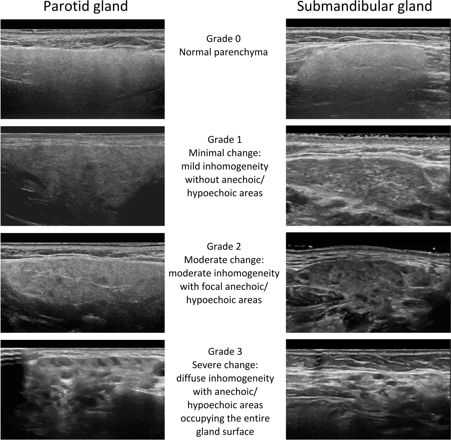
The newly developed and validated OMERACT semiquantitative Grey-scale Scoring System for SS (0–3) was applied in the study. The scores are defined as: grade 0, normal parenchyma; grade 1, mild inhomogeneity without anechoic or hypoechoic areas and hyperechogenic bands; grade 2, moderate inhomogeneity with focal anechoic or hypoechoic areas; and grade 3, severe inhomogeneity with diffuse anechoic or hypoechoic areas occupying the entire gland or a fibrous gland.14 Based on text definitions and image examples that were available in the original publications,13 14 an ultrasound atlas with four imaging examples per grade for each gland was developed by one of the participating rheumatologists with ultrasound experience (VF) and approved by all three participating rheumatologists performing and scoring the ultrasound examinations (see figure 1). The full atlas is available as online supplemental file 1. In all patients, a consensus score was obtained for all glands based on the stored still images and video clips using the atlas. The consensus scoring was done with the assessors blinded to all clinical and laboratory results.
Supplemental material

OMERACT Ultrasound Scoring System for Sjögren. Representative examples of images reflecting the written definitions of the OMERACT scoring system grade 0–3 for (left) the parotid gland and (right) the submandibular gland. The examples can also be seen in the atlas—online supplemental file 1.
Ultrasound as a minor criterion in the classification criteria
To assess the potential value of ultrasound of the salivary glands as a minor criterion in the classification criteria of pSS, 1 extra point was added to the score used in ACR/EULAR 2016 criteria, when at least one gland had an ultrasound score of 2 or 3.
Statistics
Characteristics of pSS and non-pSS were compared using t-test, χ2 test and Cochran-Armitage test of trend as appropriate. In total, six provisional ultrasound cut-offs were tested: ≥1 gland with ultrasound score 1, 2 or 3; ≥1 gland with ultrasound score 2 or 3; ≥1 gland with ultrasound score 3; ≥2 glands with ultrasound score 1, 2 or 3; ≥2 glands with ultrasound score 2 or 3; ≥2 glands with ultrasound score 3. The diagnostic performance of these ultrasonography cut-offs was examined using the 2016 ACR/EULAR classification criteria as reference standard. The number of labial biopsies that could potentially be avoided when using ultrasound was estimated.
Results
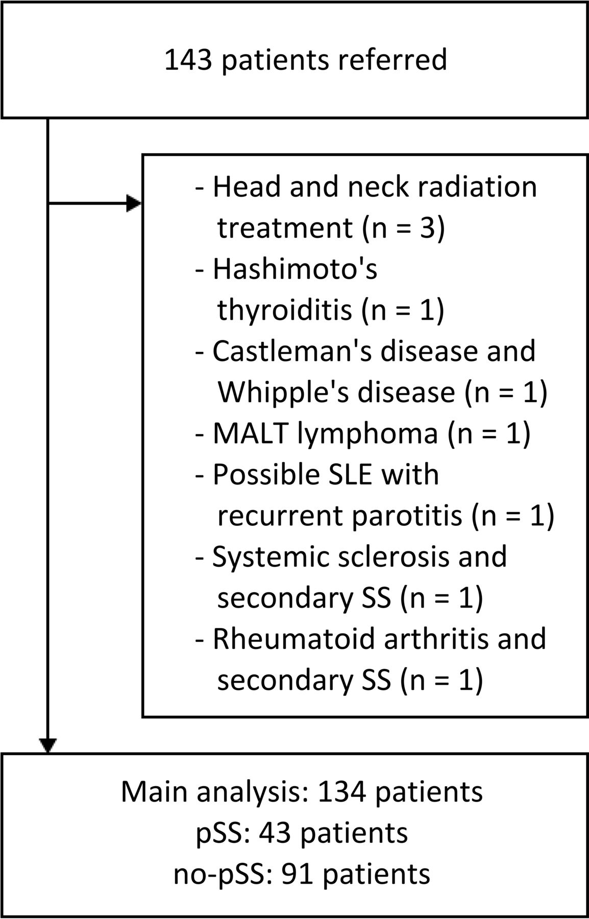
We included all 143 patients with suspected SS (sicca symptoms, ie, ocular and/or oral dryness) who were referred to our department during a 1-year period. Nine patients were excluded from the main analysis due to, for example, previous radiation therapy to the head or neck or secondary SS (see figure 2). Among the remaining 134 patients that were included in the main analysis, 43 (32%) were clinically diagnosed with pSS and all of these also fulfilled the 2016 ACR/EULAR classification criteria, while the remaining 91 (68%) patients with sicca symptoms did not receive a clinical diagnosis of pSS or fulfilled the 2016 ACR/EULAR classification criteria. Data for the ACR/EULAR classification criteria were complete, except for 27 (20%) patients that could not be formally assessed since labial biopsies were not performed: 21 patients were not offered a labial biopsy because the clinical suspicion for SS was very low, 1 patient had no labial biopsy performed due to pregnancy, while 5 patients who were recommended a biopsy did not wish to undergo the procedure. In total, 45 of the 134 (34 %) patients had a labial biopsy performed. Thirty of these patients were anti-SSA negative and 6 of these biopsies were positive.

Patient disposition. Patients with pSS: 2016 ACR/EULAR criteria for pSS were fulfilled (at least four points), all had a clinical diagnosis of pSS. Non-pSS patients: 2016 ACR/EULAR criteria for pSS not fulfilled (below 4 points, 27 patients had 1–3 points according to the 2016 ACR/EULAR criteria but did not have labial salivary gland biopsy, none of these had a clinical diagnosis of pSS). ACR, American College of Rheumatology; MALT, mucosa-associated lymphoid tissue; non-pSS, patients without pSS; pSS, primary Sjögren’s syndrome; SLE, systemic lupus erythematosus; SS, Sjögrens syndrome.
Ultrasound findings and classification criteria for pSS
Demographic data for the whole cohort and for those fulfilling and not fulfilling the ACR/EULAR classification criteria are shown in table 1. A significant difference between the two subgroups was seen for all parameters except sex and anti-CCP.
Demographics and disease characteristics for the entire cohort and separated by fulfilment of the ACR/EULAR classification criteria
Of 134 patients, 43 (32 %) patients had at least one gland with an ultrasound score 2 or 3, and 37 (28%) patients had at least two glands with an ultrasound score 2 or 3. The proportion of patients with ≥1 gland with an ultrasound score 2 or 3 was much higher in patients with pSS compared with patients without pSS (31 (72%) patients and 12 (13%) patients, respectively; p<0.001). Similarly, the proportion of patients with ≥2 glands with an ultrasound score 2 or 3 was much higher in patients with pSS compared with patients without pSS (30 (70%) patients and 7 (8 %) patients, respectively; p<0.001). The SMGs were affected slightly more frequently than the parotid glands (see table 1).
Of the 43 (32%) patients who had at least one gland with a score of 2 or 3, 35 (81%) of these patients had anti-SSA antibodies. The remaining 8 (19%) patients were negative for anti-SSA antibodies, 4 patients had a biopsy performed, of which 3 were positive and 1 was negative.
When the disease characteristics were summarised according to the highest ultrasonography score for gland pathology, see table 2, patients with a highest score of 2 or 3 among all four glands had more frequently autoantibodies, positive sialometry and positive Schirmer’s test, compared with patients who had a highest score of 0 or 1.
Disease characteristics according to highest ultrasonography score among four salivary glands
Diagnostic performance of ultrasonography findings using the 2016 ACR-EULAR pSS criteria as reference standard
We found that using a cut-off of ≥1 gland with score 2 or 3 or ≥2 glands with score 2 or 3 had a good performance for the diagnosis of pSS. These two cut-offs did not differ markedly in performance. Salivary gland ultrasound where ≥1 gland has score 2 or 3 had sensitivity 0.72 and specificity 0.91; salivary gland ultrasound where ≥2 glands had score 2 or 3 had sensitivity 0.70 and specificity 0.94 (see table 3). Also, for the positive and negative predictive values only minimal differences were found when comparing these two cut-offs (table 3).
Diagnostic performance of different US cut-offs using the 2016 ACR/EULAR primary SS criteria as reference standard
In contrast, we found that using a gland score of 1 as cut-off led to an unacceptably low specificity, while using a gland score of 3 as cut-off markedly decreased the sensitivity without a meaningful gain in specificity (see table 3).
Could biopsies potentially be avoided if ultrasound were a minor criterion in the classification criteria?
Labial biopsy and autoantibodies have the highest weight in the classification criteria. As labial biopsy is an invasive procedure that should be limited to as few patients as possible, we assessed how the patients in our cohort would fulfil the classification criteria with or without labial biopsy given a scenario where ultrasound of the major salivary glands may substitute labial biopsy in the classification criteria. When reviewing the detailed points in the classification criteria for each patient, 32 (74%) of 43 patients that were classified as pSS could be classified without ultrasound and without biopsy, that is, solely based on positive anti-SSA or anti-SSB in combination with positive Schirmer’s test and/or positive sialometry.
If ultrasonography were used as an additional item in the classification criteria, whereby at least one gland with an ultrasound score 2 or 3 would give an additional point to the criteria score, this would allow additionally 8 (6%) patients to be classified as pSS (ie, 40 out of 43 patients would have a criteria score >4) without labial biopsy being performed. Thus, the addition of ultrasound to the criteria would lead to the potential avoidance of biopsies in 8 patients. Also, 33 (25%) patients could confidently be classified as non-pSS with ultrasound and without biopsy. Overall, in this cohort 73 (54%) of the 134 patients a biopsy would not lead to a different disease classification.
Discussion
In this cross-sectional, single-centre study of 134 patients with suspected SS, we applied the OMERACT Ultrasound Grey-scale Scoring System for parenchymal changes in the large salivary glands. Glands with grade 2 and 3 were much more frequent in patients fulfilling the ACR/EULAR classification criteria than in patients who did not fulfil these criteria. Furthermore, a grade 2 or 3 in at least one gland had a high sensitivity and specificity for the diagnosis of pSS fulfilling the ACR/EULAR classification criteria and with a specificity only improving slightly when requiring pathology in at least two glands. This is partly in line with a previous study that suggested pathology in at least two glands for diagnosing pSS.13 Furthermore, a gland score of 1 was more frequent in patients without pSS than in patients with pSS and had an unacceptably low specificity for pSS, indicating that grade 1 may be considered a normal finding. A recent review paper15 addressed the large differences among published ultrasound scoring systems for salivary glands. We found that the consensus-based and validated OMERACT Grey-scale Scoring System had an improved sensitivity (0.70) compared with and an excellent specificity (0.94) in line with previously published scoring systems.9–12 Furthermore, it is encouraging that the scoring system maintains a very high specificity in this study compared with other scoring systems, since a high specificity is necessary for the use in routine care. However, a normal ultrasound cannot rule out the presence of pSS.
We chose to evaluate the ultrasound findings in relation to fulfilment of the pSS classification criteria as reference standard to avoid circularity of reasoning. We acknowledge that patients with a clinical suspicion of pSS and high ultrasound scores might be considered by some to have pSS even though they do not fulfil ACR/EULAR classification criteria.
We assessed the possible impact of ultrasound for classifying pSS if incorporated into the classification criteria5 6 16–20 with a weight of 1 point, as previously suggested.4 5 In our study, 8 (6%) patients who had 3 points in the current classification criteria would potentially gain 1 additional point by ultrasound and then fulfilling the criteria for pSS. We do not yet have long-term follow-up data available for those patients that were not diagnosed with pSS. In future studies, it should be pursued whether the ultrasound findings are predictive for developing other pSS features over time.
In the current study, we assessed to what extent biopsies could be avoided by adding ultrasound with a weight of 1 into the classification criteria. Of the 134 patients, when using clinical examination and ultrasound assessment 61 (46%) patients could avoid the invasive procedure and still be classified as pSS, thereby reducing the number of patients that needed to undergo labial biopsy to 73 (54%). Although it seems that the systematic use of ultrasonography could reduce the number of labial biopsies, it needs further testing.
Several studies have shown ultrasound of the large salivary glands to have diagnostic value for pSS,10–12 which we also demonstrated using the new OMERACT Grey-scale Scoring System when considering grade 2 and 3 as sign of pathology. Ultrasound may be taken into consideration when making a clinical diagnosis in patients with suspected pSS, if other parameters strongly indicate a clinical diagnosis of pSS, and we support the proposal of adding ultrasound into the classification. We hope that our atlas will help implement the use of ultrasound of the large salivary glands in patients suspected for pSS and ensure consistency in grading the lesions.
Strengths of this study include the application of the OMERACT consensus-based and validated Grey-scale Scoring System for SS and the development of an atlas, which ensured a uniform scoring in all patients. Furthermore, a recent study found the intra-rater and inter-rater reliability for the OMERACT Grey-scale Scoring System to be substantial to almost perfect among 20 rheumatologists.21 It might be considered a limitation that the clinicians were not blinded to the results of the ultrasound examination. However, in our data analysis we applied the ACR/EULAR classification criteria as gold standard in which ultrasound is not a part, and the potential impact of the ultrasound results to the clinician was therefore avoided.
Conclusion
The OMERACT Ultrasound Grey-scale Scoring System for SS has good sensitivity and excellent specificity for fulfilling the pSS classification criteria, when a grey-scale score of 2 or 3 in at least one gland is considered indicative of SS syndrome. Our data supports that ultrasound play an important role for diagnosing pSS and should be considered incorporated in the classification criteria. The atlas, which is available as online supplemental file 1, may be helpful in clinical practice and trials when grading lesions of the large salivary glands by ultrasound.
Data availability statement
Data are available upon reasonable request.
Ethics statements
Ethics approval
The local ethics committee evaluated the project (H-20033020) and found that an ethical approval was not needed due to the retrospective, observational design.
References
Supplementary materials
Supplementary Data
This web only file has been produced by the BMJ Publishing Group from an electronic file supplied by the author(s) and has not been edited for content.
Supplementary Data
This web only file has been produced by the BMJ Publishing Group from an electronic file supplied by the author(s) and has not been edited for content.
Footnotes
Contributors VF, UMD and LT collected data and designed the project. SK performed statistical analysis. VF, UMD, SK and LT interpreted data and wrote the manuscript. All authors have approved the final manuscript.
Funding The authors have not declared a specific grant for this research from any funding agency in the public, commercial or not-for-profit sectors.
Competing interests None declared.
Provenance and peer review Not commissioned; externally peer reviewed.