Article Text

Abstract
Objective To develop an ultrasonographic image acquisition protocol and a joint-specific scoring system for synovitis with reference atlas in patients with juvenile idiopathic arthritis (JIA) and to assess the reliability of the system.
Methods Seven rheumatologists with extensive ultrasound experience developed a scanning protocol and a semiquantitative joint-specific scoring system for B-mode (BM) synovitis for the elbow, wrist, metacarpophalangeal 2–3, proximal interphalangeal 2–3, hip, knee, ankle and metatarsophalangeal 2–3 joints. An ultrasonographic reference atlas for BM synovitis, divided in four age groups (2–4, 5–8, 9–12, 13–18 years), and power Doppler (PD) activity was then developed. Reliability was assessed for all joints on still images and in a live exercise including 10 patients with JIA, calculated by intraclass correlation coefficient (ICC) and weighted kappa.
Results A scanning protocol and scoring system for multiple joints with reference atlas composed of images with four different score levels for BM and PD were developed. Still image scoring for BM synovitis on joint level showed good to excellent intra-reader reliability (ICC/kappa ranges: 0.75–0.95/0.63–0.91) and moderate to excellent inter-reader reliability (ICC/kappa ranges: 0.89–0.99/0.50–0.91). Still image scoring for PD activity showed excellent intra-reader and inter-reader reliability (ICC/kappa: 0.96/0.91 and ICC/kappa: 0.97/0.80, respectively). In the live scoring, inter-reader reliability (ICC/kappa) was moderate to excellent for BM synovitis (0.94/0.51) and PD activity (0.91/0.60).
Conclusion An ultrasonographic image acquisition protocol and joint-specific scoring system with reference atlas were developed and demonstrated moderate to excellent reliability for scoring of synovitis in patients with JIA. This can be a valuable tool in clinical practice and future research.
- ultrasonography
- arthritis
- juvenile
- inflammation
- synovitis
Data availability statement
Data are available upon reasonable request to the corresponding author.
This is an open access article distributed in accordance with the Creative Commons Attribution Non Commercial (CC BY-NC 4.0) license, which permits others to distribute, remix, adapt, build upon this work non-commercially, and license their derivative works on different terms, provided the original work is properly cited, appropriate credit is given, any changes made indicated, and the use is non-commercial. See: http://creativecommons.org/licenses/by-nc/4.0/.
Statistics from Altmetric.com
Key messages
What is already known about this subject?
Ultrasound is an important tool in the evaluation of joint inflammation but can be difficult to interpret in children.
What does this study add?
A novel, reliable joint-specific scoring system for synovitis with reference atlas for frequently affected joints in JIA was developed.
How might this impact on clinical practice or further developments?
The combination of a defined joint-specific scoring system with reference atlas for assessing synovitis may introduce an intuitive and feasible implementation of ultrasound in patients with JIA.
Introduction
Persistent joint inflammation is the hallmark feature in juvenile idiopathic arthritis (JIA).1 If not properly reversed by treatment this inflammation can ultimately destroy the joints, explaining why JIA used to be one of the most disabling childhood diseases.1 Over the last 20 years the development of new effective drugs has improved the outcome of JIA and reduced the burden of disease for the afflicted children.2 3 Still, less than half of these patients achieve sustained inactive disease.4
The challenge of achieving inactive disease in JIA relates closely to the challenge of monitoring disease activity to detect persistent joint inflammation, and step up therapy when required. Symptoms and clinical signs of joint inflammation can be difficult to assess and interpret in children due to vague complaints and clinically challenging anatomical regions.5 6 This emphasises the need for sensitive measures of joint inflammation to assess disease activity and treatment response.7
Ultrasound is an important tool in the evaluation of joint inflammation and provides a unique possibility for systematically assessing all joints in one single bedside examination.8 9 Ultrasound is well suited for use in children, is relatively cheap and feasible, and does not require sedation or exposure to ionising radiation. However, ultrasound interpretation in children requires thorough knowledge of the age-dependent variability in the maturing skeleton.8 10–12 The Outcome Measures in Rheumatology (OMERACT) ultrasound paediatric group has started the process of standardising ultrasound assessments in children. The group has developed definitions of sonographic features of joints and descriptions of scanning approaches for the knee, ankle, wrist and second metacarpophalangeal (MCP) joints in healthy children, and preliminary ultrasound definitions of synovitis.11 13 14 However, an ultrasonographic scoring system has so far not been published by OMERACT. The use of an ultrasonographic atlas as reference for scoring of synovitis in patients with rheumatoid arthritis has shown high reliability,15 but cannot be directly applied to children due to their distinctive anatomy during growth. To our knowledge, two scoring systems in JIA exist.16 17 One, the scoring system of paediatric synovitis (PedSynS), proposes a single standard scoring system but does not clearly apply to all joints.16 The second offers joint-specific scoring for the knee but cannot be applied to other joints.17 The two scoring systems have made important contributions to standardise the use of ultrasound in patients with JIA. However, they do not fully encompass the heterogeneous joint distribution in these patients. In an effort to further broaden the application and feasibility of musculoskeletal ultrasound in JIA, we wanted to examine if a joint-specific approach and an ultrasonographic reference atlas for patients with JIA could improve the feasibility of ultrasound examination and scoring of synovitis in these patients.
The objectives of this study were to develop an ultrasonographic image acquisition protocol and a semiquantitative joint-specific scoring system with an age-divided reference atlas for scoring of synovitis in patients with JIA, and to assess the reliability of the scoring system.
Methods
The study was performed at Oslo University Hospital (OUH) at the Department of Rheumatology from January 2018 to October 2020 and conducted through the following six steps: Development of an ultrasonographic image acquisition protocol and still image collection (step 1 and 2), development of a semiquantitative joint-specific scoring system with reference atlas (step 3 and 4), reliability testing of the scoring system with reference atlas including a still image scoring (step 5) and a live exercise (step 6), (flowchart in online supplemental figure 1).
Supplemental material
Development of an image acquisition protocol
In the first step, one adult and six paediatric rheumatologists with extensive experience in musculoskeletal ultrasound (5–20 years) developed an image acquisition protocol for frequently affected joints in JIA (anterior elbow, posterior elbow, radiocarpal, midcarpal, MCP2–3 (dorsal), proximal interphalangeal (PIP) 2–3 (dorsal and volar), hip, knee (suprapatellar recess and lateral parapatellar recess), tibiotalar, talonavicular, anterior subtalar, posterior subtalar and metatarsophalangeal (MTP) 2–3 (dorsal)). Different views for some joints were chosen to provide additional information when scoring synovitis, i.e. the anterior and posterior elbow were considered to be recesses of the same joint. The protocol was built on established scanning approaches.13 15 17 18 However, since only a few scanning procedures have previously been described for children,13 17 the image acquisition protocol was adjusted and further specified to be applicable for paediatric joints through a consensus process driven by literature review, discussions and face-to-face meetings including two live exercises where seven patients with JIA (ages 3–16 years), who volunteered to participate, were assessed. The first session was held in May 2018 (NKS, A-BA, HBH, VL), the second in June 2018 (NKS, A-BA, HBH, BF, JR, VL). General aspects like defining important anatomical landmarks and the optimal position of the patient to acquire a good image were discussed. Joint-specific landmarks were included as part of the image acquisition protocol to ensure a standardised scanning position. For the wrist, the validated scanning procedure by Collado et al was applied.13 For the knee, the scanning procedure published by Ting et al was used.17 For the remaining joints, the image acquisition protocol including landmarks was developed through the consensus process described above. Finally, a protocol with specific instructions for each joint was developed and full consensus among the rheumatologists was reached.
Collection of still images
In the second step, ultrasonographic images of joints with different degrees of pathology were collected from the inpatient and outpatient rheumatology clinics at OUH according to the predefined image acquisitions. The images were collected during routine ultrasound examination as part of daily clinical practice. Two GE Logiq S8 ultrasound machines with linear probes (6–15 MHz) and hockey sticks (8–18 MHz) were used to acquire and collect the images. Approximately 5000 images were obtained and categorised jointwise in four age groups according to age-related changes; 2–4, 5–8, 9–12, 13–18 years.13 The images served as a database for the third step.
Development of an ultrasonographic scoring system and reference atlas
In the third step, the rheumatologists performed a literature review and discussed important aspects related to synovitis in different joints in patients with JIA. They also reviewed ultrasonographic images (obtained from the database of 5000 images) with different degrees of B-mode (BM) synovitis. They decided for a joint-specific scoring system because a single standard system did not clearly apply to all joints. A semiquantitative scoring system (0–3) was chosen in accordance with what has previously been done.16 17 19 Sonographic features of synovitis were defined according to the OMERACT ultrasound group.14 Based on this, the rheumatologists proposed scores for different grades and joints and discussed the suggestions on teleconferences, mail correspondence and face-to-face meetings. The nomenclature ‘mild, moderate and severe’ was included in the joint-specific scoring system to further elaborate the severity of the findings. Through this dynamic process a preliminary four-point semiquantitative joint-specific scoring system for BM synovitis, ranging from grade 0 (normal) to grade 3 (severe) for each joint was developed and full consensus reached among the rheumatologists. The scoring system for the knee was built on a newly published system that displayed good reliability, and was in accordance with our aim of developing a joint-specific scoring system.17
Scoring of Doppler activity was applied to Doppler signals detected within synovial hypertrophy (BM score >grade 0), harmonising with the joint-specific scoring system for BM synovitis, and in accordance with the definitions developed by the OMERACT ultrasound group where Doppler signals must be detected within synovial hypertrophy to be considered as a sign of synovitis.14 The scoring system for Doppler activity followed Ting et al.17 Power Doppler (PD) was chosen instead of colour Doppler because the participating rheumatologists used PD in their daily clinical practice and had more experience with this method.
In the fourth step, NKS selected BM images from the collection of 5000 images in the database that best corresponded to the scoring system for each joint in the four age ranges to establish an age-divided reference atlas for scoring of BM synovitis. All selected images were reviewed and approved by the research group. The reference atlas was finally composed of 224 characteristic BM images (14 joint regions with four different score levels in four age-groups). Representative PD images were selected by NKS from the database of 5000 images without accounting for an equal distribution of images from all age groups to develop a reference atlas for scoring of PD activity. Eventually, the reference atlas consisted of 51 distinctive PD images (13 joint regions (the hip was not included as PD signals are rarely found in this joint) with four different score levels). Due to missing images for some grades in our database, two BM and seven PD images were added to the atlas by JR working at a collaborating centre using GE Logiq E9 ultrasound machines with linear probes (6–15 MHz) or hockey sticks (8–18 MHz).
During a meeting in October 2019 the feasibility and face validity of the system with reference atlas were tested by four rheumatologists (NKS, PB, VL, JR) in a scoring exercise consisting of 69 ultrasonographic still images of joints with different degrees of pathology from patients with JIA (ages 2–18 years). The images were randomly selected by NKS from the database of 5000 images. Then a live exercise performing ultrasound of the joints included in the image acquisition protocol was done bilaterally in four patients with JIA (ages 2–15 years). The assessors were blinded to each other’s scoring and clinical information, but the patient’s age was known. The images were scored individually according to the system and saved for a following discussion concerning the scoring of the images obtained. Feasibility was assessed by the time spent performing the ultrasound examination and scoring of pathology defined to be within 30 min, and the tolerance of the examined children.
The definitions and scores were thoroughly discussed during the still image scoring and live scoring exercises. The main sources of initial disagreement concerned the development of suitable scores for the subtalar, wrist and finger joints, where the distribution of synovial hypertrophy/effusion was discussed in detail. The rheumatologists agreed on the use of percentages to differentiate grades in some joints and that the terms ‘without overall convex shape’ and ‘clearly convex shape’ could distinguish between grades 2 and 3 in the MCP, PIP and MTP joints. They also found that scoring of the knee joint (suprapatellar recess) in the youngest children could underestimate the degree of pathology due to their relatively shorter femur and made adjustments in the protocol. In case of disagreement, ultrasonographic images were reviewed and scoring feasibility discussed to reach harmonisation of the scoring definitions. During these exercises the system was adjusted, and the reference atlas improved accordingly by including images that satisfied the criteria to the scoring system. Finally, 100% consensus was reached among the rheumatologists for the scoring system and atlas.
Reliability testing of the scoring system with reference atlas on still images
The fifth step was conducted in December 2019/January 2020 where the same group (NKS, PB, VL, JR) performed an intra-reader and inter-reader reliability study. From the database of 5000 images, NKS selected 370 ultrasonographic still images of joints with different degrees of BM synovitis from patients with JIA (ages 2–18 years), consisting of at least 20 images from each joint included in the scoring system for BM synovitis. The number of images per age varied for each joint, but every joint had images from all four age groups. The images were scored jointwise with the novel scoring system and atlas for BM synovitis as reference. The rheumatologists were blinded to each other’s scoring and clinical information. The images were rearranged for a second round of scoring that was done at least 2 weeks later. To assess reliability of the scoring system and atlas for PD activity, three rheumatologists (NKS, PB, VL) scored 37 ultrasonographic still images of joints with different degrees of PD activity selected by NKS from the database of 5000 images. After 3 weeks, the images were rearranged, and a second PD scoring was performed.
Reliability testing of the scoring system with reference atlas in a live exercise
The sixth step was a live scoring exercise including 10 consecutive patients fulfilling the International League of Associations for Rheumatology (ILAR) criteria for JIA,20 attending the paediatric rheumatology clinic at OUH in the period September/October 2020. Signed informed consent was obtained by patients and/or parents. Each patient was assessed bilaterally by three rheumatologists (NKS, PB, VL) within 1–2 days, performing ultrasound of the joints included in the image acquisition protocol using a GE Logiq S8 machine with linear probe (6–15 MHz) and standardised settings for BM and PD (pulse repetition frequency 0.6 kHz, frequency 7.7 MHz and low wall filter). The rheumatologists were blinded to each other’s scoring and clinical information and had digital and printed versions of the scoring system and atlas available during the examinations. The assessed joints were used to derive separate BM synovitis and PD activity sum scores. For the joints assessed from two views, one view was selected to avoid increased weighting of these joints. The ultrasound sum score included the anterior elbow, radiocarpal, midcarpal, MCP2–3 dorsal, PIP2–3 volar, hip, knee (suprapatellar recess), tibiotalar, talonavicular, anterior subtalar and MTP2–3 dorsal joints.
Patient and public involvement
Patients have not actively participated in the planning of this study. However, the patients included in the study and the Norwegian Rheumatism Association have endorsed the project and voiced that this is of true interest to the patient community, especially concerning the national and international disparity in the use of ultrasound. Study results will be disseminated to patients and the public through the patient organisation’s website and newsletter.
Statistics
Descriptive statistics were calculated as mean (range) and median (range), as appropriate. Reliability testing in the still image exercise was performed on a joint level for BM scoring, and on all joints included in the scoring exercise combined for PD scoring. In the live exercise, separate ultrasound sum scores for BM synovitis and PD activity were calculated and used for reliability testing. Reliability was calculated by intraclass correlation coefficient (ICC, absolute-agreement, two-way mixed-effects model) and weighted kappa with linear weights.21 Intra-reader reliability was calculated as single measure (sm) ICC and Cohen’s weighted kappa, reported as mean (SD) between readers. Inter-reader reliability was calculated as average measure (avm) ICC, with 95% CI and Light’s weighted kappa (SD). ICC and kappa values 0.2–0.4 were considered fair, 0.41–0.6 moderate, 0.61–0.8 good and >0.81 excellent. Analyses were performed using SPSS V.27.
Results
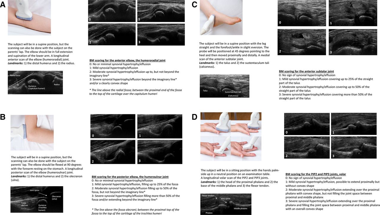
An ultrasonographic image acquisition protocol for frequently affected joints in JIA was first established (table 1). A semiquantitative joint-specific scoring system (table 2) with an age-divided reference atlas for scoring of BM synovitis and a reference atlas for scoring of PD activity in patients with JIA was then developed. The atlas is shown with example illustrations in figure 1A-D and figure 2. For the full scoring system and atlas see online supplementary file. Feasibility assessment showed that the time spent on the ultrasound examination and scoring of pathology was attainable within 30 min and was well tolerated by the participants.
Supplemental material
Supplemental material
Supplemental material
Supplemental material
Supplemental material
Ultrasonographic image acquisition protocol for frequently affected joints in juvenile idiopathic arthritis
Ultrasonographic semiquantitative joint-specific scoring system for BM in juvenile idiopathic arthritis

(A–D) Description of ultrasound examination and scoring of B-mode (BM) synovitis from the ultrasonographic BM reference atlas in juvenile idiopathic arthritis (JIA). (A) The elbow joint, longitudinal anterior scan (2–4 years). (B) The elbow joint, longitudinal posterior scan (5–8 years). (C) The anterior subtalar joint, medial scan (9–12 years). (D) The proximal interphalangeal (PIP)2 and PIP3 joints, longitudinal volar scan (13–18 years).

Description of ultrasound examination and scoring of power Doppler (PD) activity for the wrist; radiocarpal and midcarpal joints (longitudinal dorsal scan) from the ultrasonographic PD reference atlas in juvenile idiopathic arthritis (JIA).
Reliability for BM synovitis scoring on still images
Intra-reader reliability for BM synovitis scoring on still images on a joint level was good to excellent (smICC range 0.75–0.95 and weighted kappa range 0.63–0.91). Inter-reader reliability for BM synovitis scoring on still images, assessed by avmICC (range) for each joint was excellent (0.89–0.99) and weighted kappa (range) was moderate to excellent (0.50–0.91). Jointwise results for the BM synovitis still image scoring are presented in table 3.
Intra-reader and inter-reader reliability for B-mode (BM) synovitis scoring on still images in juvenile idiopathic arthritis (JIA)
Reliability for PD activity scoring on still images
Intra-reader reliability for PD activity scoring on still images from all joints included in the scoring exercise combined was excellent (smICC (SD) 0.96 (0.03) and weighted kappa (SD) 0.91 (0.06)). Inter-reader reliability for PD activity scoring on still images from all joints included in the scoring exercise combined, was good to excellent (avmICC (95% CI) 0.97 (0.94 to 0.98) and weighted kappa (SD) 0.80 (0.06)).
Reliability for BM synovitis and PD activity scoring in live exercise
Ten patients, seven girls and three boys were included in the live scoring exercise with a median age (range) 7.5 years (3–10 years). Seven patients had oligoarthritis (70%), three patients had rheumatoid factor-negative polyarthritis (30%). Median disease duration (range) was 10 months (0–65 months). Five patients were treated with methotrexate, one with etanercept, one with nonsteroidal anti-inflammatory drug (NSAID)s and three patients were without systemic treatment. The number of patients assessed in each age group was two in group 1 (ages 2–4 years), five in group 2 (ages 5–8 years) and three in group 3 (ages 9–12 years). Ultrasound findings of a BM synovitis score ≥1 were present in 29 of 280 joint regions (10.4%) and a PD activity score ≥1 in 13 of the 29 joints with BM synovitis (44.8%). The most frequently affected joints (number) were the knee (9), anterior elbow (5) and anterior subtalar (3) for BM synovitis, and the radiocarpal (4) and knee (3) for PD activity. Mean (range) ultrasound sum scores for BM synovitis and PD activity were 7.5 (5.8–9.6) and 2.2 (1.8–2.9), respectively. Inter-reader reliability (avmICC (95% CI)) for BM synovitis and PD activity ultrasound sum scores were 0.94 (0.72 to 0.99) and 0.91 (0.74 to 0.97), respectively. Weighted kappa (SD) was 0.51 (0.09) for BM synovitis and 0.60 (0.12) for PD activity.
Discussion
For the first time we present an ultrasonographic image acquisition protocol and a semiquantitative joint-specific scoring system for synovitis with an age-divided reference atlas for BM synovitis and a reference atlas for PD activity for frequently affected joints in patients with JIA. The present study demonstrated overall moderate to excellent reliability.
The image acquisition protocol ensured a standardised ultrasound examination in the practical sessions and live scoring exercises. Some of the views were adapted and adjusted from the OMERACT ultrasound paediatric group.13 Their scanning approaches showed to be applicable in children regardless of age. In our study, the image acquisition protocol was easily learnt and highly feasible, probably because of the thorough descriptions and illustrative ultrasound images with important anatomical landmarks for each joint. As the pattern of joint involvement seems to be of prognostic importance,22 23 a standardised and systematic ultrasound examination might be able to improve assessment of disease activity and individualise treatment in patients with JIA.
The novel scoring system proposes joint-specific scores for frequently affected joints in JIA. A single standard paediatric scoring system may have some limitations in that it does not clearly apply to all joints.16 For instance, grade 2 and grade 3 BM synovitis in the PedSynS is partly defined by whether or not the joint recess is extending over the bone diaphysis.16 The score may be difficult to use for joints adjacent to short bones without diaphysial bone structures. A joint-specific scoring system for the knee in patients with JIA was recently developed and demonstrated good reliability.17 This provided the basis for our further development of joint-specific scores for frequently affected joints in JIA. The suprapatellar recess was first scored with the scoring system presented by Ting et al.17 However, in the smallest children we discovered that this system could underestimate the degree of pathology due to their relatively shorter femur. In the image acquisition protocol, we therefore added that for the youngest children the patella should fill a third of the image on the ultrasound screen when scoring for pathology.
The variable sonoanatomy in the growing child may lead to pitfalls even when performed by experienced rheumatologists, and there is a lack of published age-specific and joint-specific imaging data in the literature. The images used in this study were selected from our database consisting of approximately 5000 ultrasonographic images. These images were collected during routine ultrasound examination as part of daily clinical practice from patients with JIA attending our inpatient and outpatient clinics. We therefore believe that our selection of images is representative of the patients with JIA seen in clinical practice. The comprehensive ultrasonographic atlas consisting of 224 BM images of normal and inflamed joints divided in four age groups and 51 images with semiquantitative scores for the presence of PD activity, enables the sonographer to recognise age-specific and joint-specific ultrasonographic findings of synovitis and to score ultrasound images according to the best possible match in the reference atlas. The combination of a defined joint-specific scoring system with reference atlas for assessing synovitis may introduce an intuitive and feasible implementation of ultrasound in patients with JIA.
The still image scoring exercise demonstrated moderate to excellent reliability for all joints. At joint level, the scoring of BM synovitis in our study showed the highest ICC and kappa values for the posterior elbow, knee, tibiotalar, anterior subtalar and the MTP joints (table 3). The good reliability for the scoring of the knee was in accordance with data reported by Ting et al.17
The subtalar joint is one of the most difficult joints to assess clinically in the ankle, but this joint is often involved in patients with JIA.6 24 25 To our knowledge, image acquisitions and scoring systems for the paediatric anterior and posterior subtalar joints have not been published before. We therefore included them in our scanning protocol and scoring system, which is also in accordance with suggestions by others.25 In our study, the joint-specific scoring with illustrative images in the reference atlas of the anterior and posterior subtalar joints showed good to excellent reliability.
Interpretation of PD signals in children is complicated due to a variable degree of physiological blood flow within the joint that can easily be misinterpreted as inflammation. The OMERACT ultrasound group has started the process of defining age-related vascularisation of joints in healthy children,13 26 and developed preliminary definitions of synovitis in children which define that Doppler signals must be detected within synovial hypertrophy to be considered as a sign of synovitis.14 Our reference atlas for scoring of PD activity might improve the feasibility of this ultrasonographic feature, but further studies are needed regarding the detection of abnormal vascularisation in the paediatric joint.
Inter-reader reliability on live scoring has only been reported in few JIA studies. The live scoring of 10 patients with JIA in this study demonstrated good reliability, which is comparable to the results presented by Magni-Manzoni et al.19
Limitations of this study are the low number of patients included in the live scoring exercise, that only three sonographers participated and that they only scanned the patients once. At the time of our live scoring exercise, we experienced that the COVID-19 situation made it impossible to conduct a large scoring exercise including more patients and readers. However, we wished to test the scoring system in a live setting and made adaptations to our project within these limitations. Previous studies have shown that inclusion of 10 patients may yield sufficient power for reliability testing.15 27 We found it feasible for three dedicated rheumatologists to do a live scoring exercise implemented in our daily clinical practice by consecutively including 10 patients with JIA disease flare in need of hospital admission. In this setting, the inter-reader reliability was good, suggesting that the scoring system with atlas is a reliable tool. Examination of potential variability in the reliability of the scoring system with respect to age or disease activity was beyond the scope of this study and should be addressed in future research.
Another limitation is the lack of comparison with healthy subjects. The main target of this study was to develop an ultrasonographic scoring system with reference atlas for patients with JIA and to test the reliability of the system. The study was not designed to compare ultrasonographic findings in healthy children with patients with JIA. However, results from available musculoskeletal ultrasound studies highlighting findings in healthy children were taken into account in the process.11–13 26 A comparison of ultrasonographic findings in healthy children with patients with JIA according to the presented scoring system could be a future study of interest.
Other limitations are that we did not have images of all grades in the atlas, and that the reference atlas for scoring of PD activity was not age-divided. However, the main goal for the PD reference atlas is to illustrate different grades of PD signals for each joint and not the age variability. Furthermore, in accordance with the definitions developed by the OMERACT ultrasound group,14 PD signals must be detected within synovial hypertrophy to be considered as a sign of synovitis, which will be clearly identified first by using the scoring system and age-divided atlas for BM synovitis as reference. In addition, we will continuously include images in our database and aspire to include the best possible reference images for all grades in the reference atlas.
The strengths of the study are the unique collection of ultrasonographic images with different degrees of pathology in four age ranges and the approach to define individual scores for a substantial number of joints in patients with JIA.
In conclusion, this study presents an ultrasonographic image acquisition protocol and a semiquantitative joint-specific scoring system for synovitis with reference atlas in patients with JIA. The study demonstrated moderate to excellent reliability when used in assessments on still images as well as on patients. We expect that the system can be a valuable tool for clinicians and future research. Future studies are needed for further validation of the scoring system with atlas, such as association to clinical measures of disease activity and the system’s sensitivity to change.
Data availability statement
Data are available upon reasonable request to the corresponding author.
Ethics statements
Ethics approval
The study was done in accordance with the Declaration of Helsinki and was approved by the Norwegian Regional Committee for Medical and Health Research Ethics (REK 2018/805) and the Data Protection Officer at Oslo University Hospital, Norway (18/11742 and 18/12493). Image storage was approved by the Data Protection Officer at OUH (18/11742).
Acknowledgments
The authors thank David Swanson, Researcher (statistician), PhD, for statistical advice, and Øystein Horgmo, senior photographer, for taking photographs of probe positioning to the ultrasonographic atlas. The authors also thank all the patients and their families who contributed to this study.
References
Supplementary materials
Supplementary Data
This web only file has been produced by the BMJ Publishing Group from an electronic file supplied by the author(s) and has not been edited for content.
Footnotes
Contributors NKS designed the study, made substantial contributions to acquisition, analysis and interpretation of data and writing the manuscript. PB and VL designed the study, and made substantial contributions to acquisition, interpretation of data and writing the manuscript. JR participated in the study design, made substantial contributions to acquisition, interpretation of data and writing the manuscript. A-BA, HBH and BF participated in the study design and made substantial contributions to data interpretation and writing the manuscript.
Funding The authors have not declared a specific grant for this research from any funding agency in the public, commercial or not-for-profit sectors.
Competing interests HBH reports personal fees from Lilly, personal fees from Novartis, personal fees from AbbVie, personal fees from Roche, outside the submitted work.
Provenance and peer review Not commissioned; externally peer reviewed.