Article Text

Abstract
Objective To identify clusters of peripheral involvement according to the specific location of peripheral manifestations (ie, arthritis, enthesitis and dactylitis) in patients with spondyloarthritis (SpA) including psoriatic arthritis (PsA), and to evaluate whether these clusters correspond with the clinical diagnosis of a rheumatologist.
Methods Cross-sectional study with 24 participating countries. Consecutive patients diagnosed by their rheumatologist as PsA, axial SpA or peripheral SpA were enrolled. Four different cluster analyses were conducted: one using information on the specific location from all the peripheral manifestations, and a cluster analysis for each peripheral manifestation, separately. Multiple correspondence analyses and k-means clustering methods were used. Distribution of peripheral manifestations and clinical characteristics were compared across the different clusters.
Results The different cluster analyses performed in the 4465 patients clearly distinguished a predominantly axial phenotype (cluster 1) and a predominantly peripheral phenotype (cluster 2). In the predominantly axial phenotype, hip involvement and lower limb large joint arthritis, heel enthesitis and lack of dactylitis were more prevalent. In the predominantly peripheral phenotype, different subgroups were distinguished based on the type and location of peripheral involvement: a predominantly involvement of upper versus lower limbs joints, a predominantly axial enthesitis versus peripheral enthesitis, and predominantly finger versus toe involvement in dactylitis. A poor agreement between the clusters and the rheumatologist‘s diagnosis as well as with the classification criteria was found.
Conclusion These results suggest the presence of two main phenotypes (predominantly axial and predominantly peripheral) based on the presence and location of the peripheral manifestations.
- spondylitis
- ankylosing
- arthritis
- psoriatic
- arthritis
Data availability statement
Data are available upon reasonable request. Data are available on reasonable request. Researchers willing to use data collected during the study should contact the first author, who will send a study proposal template to be completed by the applicant. Thereafter, the steering committee of the ASAS-PerSpA study will approve (or not) the proposal and proceed to the data sharing.
This is an open access article distributed in accordance with the Creative Commons Attribution Non Commercial (CC BY-NC 4.0) license, which permits others to distribute, remix, adapt, build upon this work non-commercially, and license their derivative works on different terms, provided the original work is properly cited, appropriate credit is given, any changes made indicated, and the use is non-commercial. See: http://creativecommons.org/licenses/by-nc/4.0/.
Statistics from Altmetric.com
Key messages
What is already known about this subject?
Peripheral manifestations can occur across the entire spectrum of spondyloarthritis (SpA) and they play a key role in deciding about an exact diagnosis in patients with SpA.
Recently, the worldwide Assessment of Spondyloarthritis international Society-peripheral SpA (PerSpA) study demonstrated that all different peripheral features can be found in all subtypes of SpA (including psoriatic arthritis (PsA)), and that differences are quantitative rather than qualitative.
However, these differences in frequency and distribution may influence the clinical diagnosis by the rheumatologist, based on their own experience or knowledge.
What does this study add?
These results distinguish two main phenotypes (a predominantly axial and a predominantly peripheral phenotype) on the basis of the peripheral manifestations, which fitted a clinical diagnosis of axial SpA (axSpA) and PerSpA (including PsA), respectively.
Key messages
How might this impact on clinical practice or future developments?
The often-artificial difference between axSpA and PerSpA therefore continues to make sense, although an important overlap of peripheral manifestations was found across the different underlying entities.
These results also confirm the variability of making a diagnosis by rheumatologists depending on the presence of peripheral manifestations.
Introduction
Spondyloarthritis (SpA) represents a chronic inflammatory disease that typically affects the axial skeleton and sacroiliac joints.1 Nevertheless, SpA also often affects the appendicular skeleton with peripheral manifestations such as arthritis, enthesitis and dactylitis. In 2009, the Assessment of Spondyloarthritis international Society (ASAS) introduced the concept of axial (axSpA) and peripheral SpA (pSpA) for patients with predominant axial or peripheral symptoms, respectively.2 In parallel, the CASPAR Study Group proposed specific criteria for the classification of psoriatic arthritis (PsA).3 Whether PsA can be considered part of SpA or as a separate entity has been a matter of debate in the last few years, since many similarities and differences have been described between these two diseases: first, inflammatory back pain (IBP) and radiographic sacroiliitis can appear in both groups.4 Second, both patients with PsA and SpA exhibit peripheral musculoskeletal manifestations, but a higher prevalence of such manifestations (especially dactylitis) and a lower prevalence of HLA-B27 positivity have been reported in patients with PsA.5 Moreover, the typical clinical presentation of articular involvement in pSpA has been described as an asymmetric, monoarticular or oligoarticular inflammatory arthritis that involves the lower limbs more frequently than the upper limbs,6 while a variety of arthritis patterns are seen in PsA. It is still under debate whether polyarticular inflammatory arthritis, which often involves the distal interphalangeal joints (a typical clinical presentation in patients with PsA) should be seen as part of pSpA.7
One major concern about studies that compare these subtypes is the fact that they usually differentiate patients either as SpA or PsA depending on the rheumatologist’s opinion, which in many cases is influenced by their education and background in the field. As an example, a patient with radiographic sacroiliitis and cutaneous psoriasis may be diagnosed as ‘axSpA with psoriasis’ or ‘axial PsA’ depending on the rheumatologist’s preferences.
Recently, the worldwide ASAS-PerSpA study demonstrated that all different peripheral features can be found in all subtypes of SpA (including PsA), and that differences are quantitative rather than qualitative.8 However, as explained above, these differences in frequency and distribution may influence the clinical diagnosis by the rheumatologist, based on their own experience or knowledge. This clinical reasoning results in the diagnosis of either axSpA, pSpA or PsA. The use of exploratory analysis for grouping patients (clusters) from the whole spectrum of SpA may be useful to group patients according to their clinical characteristics, and to confirm whether certain clusters are more frequent in patients with a specified diagnosis (either axSpA, pSpA or PsA) according to the rheumatologist. Clustering analysis represents a class of unsupervised exploratory analytical techniques, which aims to identify homogeneous groups of cases without taking prior information about the group of cluster or membership into account. In this sense, this technique may be especially valuable in the identification of clusters with regard to the peripheral involvement in the whole spectrum of SpA.
In this study, we conducted an unsupervised analysis with the aim to identify clusters of peripheral involvement based on the specific location of these manifestations in the whole spectrum of SpA, and to evaluate whether these clusters are in agreement with the rheumatologist’s diagnosis (ie, axSpA, pSpA and PsA) and classification criteria.
Methods
Patients and study design
The ASAS-PerSpA is an observational, cross-sectional, multicentre and international study with 24 participating countries and 68 centres from four continents (Africa, America, Asia and Europe). Consecutive adult patients (eg, at least 18 years old) with a diagnosis of SpA (either axSpA, pSpA or PsA) according to clinical diagnosis of their rheumatologist and who were able to understand and complete questionnaires were included. The ASAS-PerSpA study has been previously described in detail.8 Written informed consent was obtained from all subjects before enrolment.
Collected data
Five different categories of data were collected:
Demographics: age, sex, smoking and alcohol intake.
Disease characteristics: the investigators were asked to name the diagnosis that in their opinion best described the disease of the patient. They could choose from the following list: axSpA, PsA, pSpA, inflammatory bowel disease (IBD)-SpA, ReA, Juv-SpA, or they could name another disease. In addition, information about HLA-B27 status, axial involvement according to the rheumatologist, uveitis, psoriasis and IBD were collected. The individual items from the ASAS axSpA, ASAS pSpA and CASPAR criteria were collected.2 3 However, pSpA ASAS criteria were evaluated in two ways: (a) following the strict rules considering IBP as excluding item for ASAS pSpA criteria (‘strict criteria’) and (b) ignoring IBP as excluding item (‘non-strict criteria’). The use of conventional synthetic and biological disease-modifying antirheumatic drugs was also collected.
Peripheral musculoskeletal manifestations: history or current presence of specific locations of arthritis (including hip and shoulder involvement), enthesitis and dactylitis that the patient has ever suffered from in the course of their disease were collected.
Disease activity and disease burden: current disease activity was measured by the Bath Ankylosing Spondylitis Disease Activity Index (BASDAI),9 and the Ankylosing Spondylitis Disease Activity Score was calculated with the C reactive protein (ASDAS-CRP).10 The Bath Ankylosing Spondylitis Functional Index (BASFI) and the ASAS Health Index (ASAS-HI) were used to evaluate physical function and functioning and health, respectively.11 12 Finally, the self-reported Fibromyalgia Rapid Screening Tool (FiRST) was applied to determine the presence of concomitant fibromyalgia.13
All information was obtained by a study investigator or research nurse during a face-to-face interview at a study visit, which included a review of the medical record. A centralised electronic case report form was used.
Statistical analysis
Unsupervised statistical learning methods were used to discover inherent but hidden patterns in the data. More specifically, for peripheral musculoskeletal manifestations (ie, arthritis, enthesitis and dactylitis), we aimed to split the observations into a number of subsets determined by the specific location of these manifestations at any time during the course of the disease. Four different cluster analyses were conducted: the first one using information from all peripheral musculoskeletal manifestations (ie, arthritis, enthesitis and dactylitis) with the aim to evaluate the number of patterns taking into account overall peripheral manifestations. After that, we conducted one cluster analysis for each peripheral manifestation (ie, one analysis for arthritis; a second analysis for enthesitis and a third analysis for dactylitis) to evaluate whether the patterns are different depending on the specific manifestation.
First, for each location of each peripheral manifestation, a variable was created to reflect the presence (=1) or absence of current or past involvement (=0); locations with missing information were considered as absence of involvement. Multiple correspondence analyses (MCA) were used to analyse the patterns of relationships between those variables, with plots allowing visualisation of the distance between the categories, and thus facilitating our interpretation of the data. A final characteristic of an MCA is that it is weighted, with each variable being allocated a weight that increases with its scarcity, in order to make any differences relating to the rarest and most distinctive modalities especially visible. Statistically comparable individuals (and characteristics shared by these individuals) are represented on these graphs as points which tend to group together, while dissimilarity, on the other hand, results in distance.
Then, clustering was conducted using an iterative partitioning k-means method. The optimal number of clusters was estimated using the ‘NbClust’ package, which provides 30 indices and proposes the best clustering scheme from the different results obtained by varying all combinations of the number of clusters, distance measures and clustering methods.14 The NbClust provides the number of clusters proposed by all the indices and proposes the best number of clusters according to the majority rule.
After the extraction of the clusters, the distribution of the different locations of peripheral manifestations across groups was described. Sociodemographic and clinical characteristics, rheumatologist’s diagnosis, classification criteria (including ‘strict’ and ‘non-strict’ pSpA ASAS criteria), disease activity and disease burden were compared across the different clusters using the χ2 test or the exact Fisher’s test for qualitative variables, and analysis of variance or Kruskal-Wallis tests for continuous variables depending on the normality of the variables. This process was done first for all peripheral manifestations, and thereafter for each of the peripheral manifestations separately (ie, arthritis, enthesitis and dactylitis).
Data were processed and analysed using RStudio V.1.0.143.
Results
Cluster analysis on all peripheral musculoskeletal manifestations together
All the patients participating in the PerSpA were included in this analysis (n=4465). MCA using information concerning the specific location of all peripheral musculoskeletal manifestations (ie, arthritis, enthesitis and dactylitis) in the overall population is represented in online supplemental figure S1. The cloud of points (ie, variables) showed a group of variables reflecting the absence of any peripheral manifestation on the intersection of the two dimensions. Hip involvement was positioned very close (ie, associated) to this group, as well as knee, shoulder and ankle involvement. Interestingly, plantar fascia and Achilles tendon involvement was also positioned close to the group of variables which representing the absence of any peripheral manifestation. Variables representing toes and fingers involvement (either as arthritis or dactylitis) formed a conglomerate on the right bottom of the figure. Finally, a wide variety of enthesitis locations represented another conglomerate on the right top of the figure.
Supplemental material

Distribution of the location of peripheral involvement across clusters with regard to the all the peripheral musculoskeletal manifestations. DIP, distal interphalangeal joint; PIP, proximal interphalangeal joint; MTP, metatarsophalangeal; MCP, metacarpophalangeal.
The clustering algorithm and the analysis of the optimal number of clusters led to the classification of the 4465 patients into two groups (with 3984 and 481 patients each). The distribution of peripheral involvement across the two clusters is represented in figure 1. Overall, cluster 1 (that we decided to call ‘predominantly axial’ cluster) showed a low prevalence of all the peripheral manifestations in comparison with cluster 2 (that we decided to call ‘predominantly peripheral’); however, when peripheral involvement was present in the ‘predominantly axial’ cluster, it was mostly represented by hip, knee and ankle arthritis, as well as Achilles tendon and plantar fascia enthesitis. The ‘predominantly peripheral’ cluster showed a high prevalence of all the peripheral manifestations, especially arthritis.
Sociodemographic data, clinical characteristics, disease activity and disease burden across the clusters are represented in table 1. Patients from the ‘predominantly axial’ cluster (cluster 1) showed a higher prevalence of males (63.1% vs 43.5%), HLA-B27 positivity (68.6% vs 37.8%), axial involvement according to the rheumatologist (79.7% vs 52.4%) and uveitis (17.9% vs 9.8%). In addition, patients from the ‘predominantly axial’ cluster had more frequently a diagnosis of axSpA according to the rheumatologist (65.7% vs 21.2%), fulfilled more frequently axial ASAS criteria, and showed a lower burden of disease measured by the BASFI (2.9 vs 4.0) and the ASAS-HI (4.5 vs 4.9). On the other hand, patients from the ‘predominantly peripheral’ cluster (cluster 2) showed, in comparison with the other cluster, a higher prevalence of psoriasis (63.0% vs 24.5%), a higher disease activity (BASDAI 4.9 vs 3.7 and ASDAS 2.9 vs 2.5, respectively), a more frequent diagnosis of PsA (61.3% vs 18.5%), and they fulfilled more frequently the ‘strict’ peripheral ASAS (25.8% vs 10.8%), the ‘non-strict’ peripheral ASAS (46.8% vs 26.3%) and the CASPAR criteria (56.8% vs 19.3%).
Sociodemographic data, clinical characteristics, disease activity and disease burden across the two clusters with regard to the location of all the peripheral musculoskeletal manifestations
Cluster analysis on arthritis only
Online supplemental figure S2 represents the MCA using information only concerning the specific location of arthritis. The cloud of variables showed, similarly to the previous analysis, a group of variables coded 0 (ie, ‘absence’ of arthritis) on the intersection of the two dimensions. Hip involvement was positioned very close to this group, as well as knee, shoulder and ankle arthritis. Interestingly, two additional conglomerates of variables were found: one reflecting hand involvement (ie, metacarpophalangeal, and finger proximal interphalangeal and distal interphalangeal involvement of the fingers) and one reflecting feet involvement (ie, metatarsophalangeal, and toe proximal and distal interphalangeal involvement).
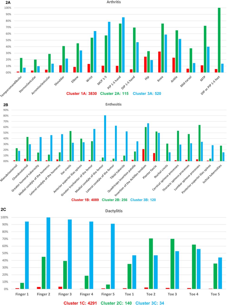
A total of 3 clusters were found, with 3839, 115 and 520 patients respectively. One cluster (1A) showed a lower prevalence of peripheral joint involvement in general than the other two clusters (figure 2A); however, in case of arthritis, it was more frequently located on the lower limbs. The other two clusters (2A and 3A) showed a high prevalence of arthritis, although predominantly in the lower limbs versus in the upper limbs, respectively.

Distribution of the location of peripheral involvement across clusters with regard to the arthritis (A), enthesitis (B) and dactylitis (C). DIP, distal interphalangeal joint; PIP, proximal interphalangeal joint; MTP, metatarsophalangeal; MCP, metacarpophalangeal.
With regard to other clinical data (table 2), cluster 1A showed a higher prevalence of males (63.9% vs 51.3% vs 41.5% for A1, A2 and A3, respectively), HLA-B27 positivity (79.8% vs 48.2% vs 34.8%), axial involvement according to the rheumatologist (80.9% vs 54.8% vs 51.5%), a lower presence of psoriasis (23.2% vs 67.8% vs 59.6%), and a more frequent diagnosis of axSpA (67.2% vs 20.9% vs 23.7%) and fulfilment of the axial ASAS criteria (70.6% vs 48.7% vs 37.1%) in comparison with clusters 2A and 3A, respectively. Conversely, clusters 2A and 3A showed a higher prevalence of enthesitis (64.3% vs 52.9% vs 42.7%, for A2 vs A3 vs A1, respectively) and dactylitis (47.8% vs 34.4% vs 11.8%) in comparison with cluster 1A, a more frequent diagnosis of PsA (61.7% vs 58.5% vs 17.2%), fulfilment of the CASPAR (63.5% vs 52.9% vs 18.1%), pSpA ‘strict’ criteria (22.6% vs 26.2% vs 10.2%) and pSpA ‘non-strict’ criteria (40.9% vs 50.4% vs 25.1%). Interestingly, these two clusters showed a higher burden of disease (BASFI 4.1 vs 4.0 vs 2.8 and ASAS-HI 9.0 vs 8.2 vs 6.3).
Sociodemographic data, clinical characteristics, disease activity and disease burden across the three clusters with regard to the location of the arthritis
The most important differences found between clusters 2A and 3A were the prevalence of uveitis (15.7% vs 8.7%) and IBD (3.5% vs 6.7%), respectively.
Cluster analysis on enthesitis only
MCA using information concerning the specific location of enthesitis is represented in online supplemental figure S3. Achilles tendon and plantar fascia involvement were positioned very close to the group of variables reflecting the absence of enthesitis. Interestingly, two other groups of variables were found: one group including axial locations (ie, chondrosternal, iliac spine, cervical and lumbar processes) and a second group including peripheral locations (femur and humeral condyles, tibial tubercle and the quadriceps patellar insertion). Three clusters were found with regard to the enthesitis location (with 4089, 256 and 120 patients, respectively). Cluster 1B showed a very low prevalence of enthesitis (figure 2B) but, if present, they were more frequently located at the Achilles tendon and plantar fascia (ie, heel enthesitis). Clusters 2B and 3B showed a high prevalence of enthesitis, with a predominant involvement of axial locations in cluster 2B and peripheral locations in cluster 3B.
Clinical data showed some similarities between clusters 1B and 2B, and differences with regard to cluster 3B (table 3), such as a higher prevalence of HLA-B27+ (66.7% vs 61.5% vs 45.5% for B1 vs B2 vs B3, respectively), axial involvement (76.4% vs 86.3% vs 68.3%), uveitis (17.2% vs 18.8% vs 9.2%) and a more frequent diagnosis of axSpA according to the rheumatologist (61.4% vs 64.1% vs 37.5%). In addition, cluster number 1B showed the lowest disease activity (BASDAI 3.7 vs 5.0 vs 5.3 and ASDAS 2.5 vs 3.1 vs 3.1) and the mildest burden of disease (BASFI 2.9 vs 4.2 vs 4.3 and ASAS-HI 6.3 vs 8.9 vs 10.4). On the other hand, cluster number 3B (the cluster with predominant peripheral locations), showed a higher prevalence of arthritis and dactylitis in comparison with clusters 1B and 2B, psoriasis (48.3% vs 28.4% vs 23.4%, respectively) and diagnosis of PsA (40.8% vs 21.5% vs 22.7%).
Sociodemographic data, clinical characteristics, disease activity and disease burden across the three clusters with regard to the location of the enthesitis
Cluster analysis on dactylitis only
MCA using information about dactylitis (online supplemental figure S4) exhibited three groups of variables: one group reflecting the absence of dactylitis, a second group with the variables referring to the fingers, and a third one with the toes. In this line, we found three clusters (4291, 140 and 34 patients, respectively) which showed a very low prevalence of dactylitis, a predominantly toes and predominantly fingers involvement, respectively (figure 2C). Cluster 1C showed, in comparison with clusters 2C and 3C, a higher prevalence of HLA-B27 positivity (66.6% vs 52.5% vs 50.0%), axial involvement (77.7% vs 54.3% vs 52.9%), a diagnosis of axial SpA according to the rheumatologist (62.6% vs 20.0% vs 11.8%) and a lower prevalence of psoriasis (26.9% vs 68.6% vs 79.4%) (table 4). On the other hand, clusters 2C and 3C showed a higher prevalence of peripheral involvement (ie, arthritis and enthesitis) and a diagnosis of PsA (65.7% vs 52.9% vs 21.5%) than cluster 1C.
Sociodemographic data, clinical characteristics, disease activity and disease burden across the three clusters with regard to the location of the dactylitis
Discussion
In this analysis conducted in the worldwide ASAS-PerSpA study,8 we tried to define groups of peripheral manifestations (ie, clusters), in a population of patients with SpA. By classifying the patients by their clinical presentation, a predominantly axial and a predominantly peripheral cluster were identified, which were mor frequently recognised by physicians as axSpA and as pSpA or PsA, respectively. Peripheral features were found in all the groups, but differences were quantitative rather than qualitative, suggesting that SpA constitutes one entity in which peripheral and axial manifestations coincides. This gives us the impression that rheumatologists worldwide consciously or subconsciously recognise these distributions on the basis of which they decide that a patient has SpA.
Two main clusters with regard to the location of peripheral musculoskeletal manifestations (ie, arthritis, enthesitis and dactylitis) were found: a ‘predominantly axial’ and a ‘predominantly peripheral’, which showed a high prevalence of clinical diagnosis of axSpA and PsA or pSpA, respectively. Typical features associated with axSpA were found in the ‘predominantly axial’ cluster: a higher prevalence of males, HLA-B27 positivity, axial involvement, uveitis and a lower prevalence of psoriasis; indeed, the diagnosis of axSpA was present in 67.5% of these patients. On the other hand, typical features associated with the peripheral presentations of SpA (either pSpA or PsA) were found in the ‘predominantly peripheral’ cluster: a higher prevalence of females, HLA-B27 negativity, a lower prevalence of axial involvement and a high prevalence of psoriasis, and a diagnosis of pSpA or PsA in these patients (11% and 61.3%, respectively). In addition, in this peripheral phenotype, 56.8% of patients fulfilled the CASPAR criteria, and only 25.8% fulfilled the ASAS peripheral criteria. The reason of this low prevalence of ASAS peripheral criteria resides in the fact that we can only apply these criteria in patients with current peripheral involvement and without current IBP,2 making both ASAS peripheral and ASAS axial criteria mutually exclusive. Indeed, ignoring current IBP as an exclusion criterion for ASAS peripheral, we found that 46.8% would fulfil these classification criteria, suggesting a high number of patients with predominantly peripheral symptoms in conjunction with IBP. This significant difference between the ‘strict’ and ‘non-strict’ ASAS peripheral criteria may be considered relevant enough to take into consideration for future adaptations of the current classification criteria.
Apart from these differences, both clusters also showed important overlaps. Both clusters (ie, ‘predominantly axial’ and ‘predominantly peripheral’) exhibited, to some extent, peripheral involvement, with the knee and the ankle among the most frequent joints affected in both groups, as well as heel enthesitis, confirming that differences in peripheral symptoms in SpA are rather quantitative than qualitative. These results are in line with a previous study using a latent class analysis (without preassumptions on the contribution of each SpA feature) that identified the clinical entity ‘axSpA with peripheral signs’, which included patients with concomitant IBP and peripheral features.15 However, in comparison with our study, patients from the ‘axSpA with peripheral signs’ showed a higher prevalence of ASAS peripheral criteria fulfilment. This is because authors applied the ASAS peripheral criteria in patients with IBP to better understand the possible overlap between pSpA and axSpA with peripheral signs.
Our results also confirm the arthritis of the large joints of the lower limbs and the heel enthesitis as typical SpA manifestations in both the axial and peripheral phenotypes.16 A remarkable finding is the high proportion of patients from the peripheral phenotype showing upper limbs involvement, represented by arthritis of the wrists, metacarpophalangeal and proximal interphalangeal joints. Although upper limbs and small joints involvement is not a typical manifestation in patients with SpA, Moll and Wright described a polyarticular subtype of PsA represented by arthritis of five or more symmetric joints resembling rheumatoid arthritis.16 17 Sixty-three per cent of patients from this peripheral phenotype suffered from psoriasis and 61.3% had a diagnosis of PsA, suggesting that rheumatologists recognise this pattern as suggestive of PsA.
The cluster analysis focusing on a specific clinical feature showed similar results, exhibiting a predominantly axial and a predominantly peripheral phenotype, but yielding three groups for each of the manifestation. In the analysis using information about the specific location of arthritis, hip involvement was especially noticed in the axial phenotype, confirming the association between this joint and axial disease18 . At variance, shoulder involvement was not especially noteworthy in the axial but was more predominant in the peripheral phenotype; in addition, in the MCA analysis, we found a strong association of this location with the knee and ankle, suggesting that, as opposed to the hip, the shoulder behaves mostly as a peripheral rather than an axial joint.
The results from the cluster analysis of enthesitis showed three clusters, with two of them resembled the axial phenotype: a ‘pure’ axial disease with axial involvement and heel enthesitis as the major clinical presentation, and a second cluster with predominantly axial enthesitis (eg, nuchal crests, thoracic and lumbar spinous processes, iliac tuberosities…). In fact, in both of these clusters, the most frequent diagnosis according to the rheumatologist was axSpA (61.4% and 64.1%, respectively). With regard to the enthesitis, a third cluster resembling a peripheral phenotype was found, which was associated more frequently with psoriasis. This group with peripheral polyenthesitis exhibited a higher prevalence of fibromyalgia according to the FiRST questionnaire in comparison with the clusters resembling axial phenotypes. However, whether these patients suffered from a true inflammatory polyenthesitis or a concomitant fibromyalgia cannot be distinguished in this study.
This study has some limitations and some strengths. One major limitation is the difficulty of correctly evaluating peripheral involvement that occurred before the study visit, which may be hampered by recall bias, especially in patients with a long-standing disease. Another limitation is the cross-sectional nature of the study, which precludes the evaluation of the cluster evolution over time. The main strength of this study is the large sample size and the number of participating countries providing a worldwide representation of patients from the whole spectrum of SpA and PsA. In fact, an important homogeneity in the distribution of clusters across regions was found, which justify the pooling analysis of the data (online supplemental table S1).
In summary, these results distinguish two main phenotypes (a predominantly axial and a predominantly peripheral phenotype) on the basis of the peripheral manifestations, which fitted a clinical diagnosis of axSpA and pSpA (including PsA), respectively. This study does not only suggest that clinicians include peripheral manifestations in their assessment of whether a patient has axial or peripheral SpA; it also emphasises several other interesting elements, such a close association between heel enthesitis and axial SpA and a difference between root joints (eg, shoulder is more related with the peripheral phenotype while hip is more related with the axial one). Thus, the often-artificial difference between axial and peripheral SpA therefore continues to make sense, although whether PsA can be considered part of SpA or as a separate entity cannot be answered with this study. However, an important overlap of peripheral manifestations was found across the different underlying entities. These results also confirm the variability of making a diagnosis by rheumatologists depending on the presence of peripheral manifestations.
Future prospective studies would be useful to assess the changes in phenotypes over time, and molecular and genetic studies are needed to evaluate whether these clinical phenotypes are genetically similar or different.
Data availability statement
Data are available upon reasonable request. Data are available on reasonable request. Researchers willing to use data collected during the study should contact the first author, who will send a study proposal template to be completed by the applicant. Thereafter, the steering committee of the ASAS-PerSpA study will approve (or not) the proposal and proceed to the data sharing.
Ethics statements
Patient consent for publication
Acknowledgments
We would like to thank all the collaborators who participated in the study: Hernán Maldonado Ficco (Hospital San Antonio de Padua, Rio Cuarto, Argentina), Rodolfo Pérez Alamino (Hospital Dr. Nicolás Avellaneda, Tucumán, Argentina), Emilio Buschiazzo (Hospital SeSEñor del Milagro, Salta, Argentina), Romina Calvo (Hospital Provincial Dr. José M. Cullen, Santa Fé, Aregntina), Vanesa Duarte (Clínica Monte Grande, Buenos Aires, Argentina), Maria Victoria Martire (Instituto Médico Platense, La Plata, Argentina), Diego Baenas (Hospital Privado de Córdoba, Córdoba, Argentina), Dora Pereira (Hospital Ricardo Gutiérrez, La Plata, Argentina), Adrian Salas (Consultorio Reumatológico, La Plata, Argentina), Juan Manuel Bande (Hospital General de Agudos Dr. E Tornú, Buenos Aires, Argentina), Alberto Berman (Centro Médico Privado de Tucumán, Tucumán, Argentina), Stephanie Belton (University of Alberta, Canada), María Paz Poblete (Facultad de Medicina Clínica Alemana – Universidad del Desarrollo, Santiago de Chile, Chile), Francisca Valenzuela (Facultad de Medicina Clínica Alemana – Universidad del Desarrollo, Santiago de Chile, Chile), Min Xiao (Third Affiliated Hospital of Sun Yat-Sen University, Guangzhou, China), CS Lau (Hong-Kong University, China), Ho Yin Chung (Hong-Kong University, China), Sherif Gamal (Cairo University, Cairo, Egypt), Catherine Lebourlout (Cochin Hospital, Paris, France), Daniel Wedling (CHU Besançon, Besançon, France), Clément Prati (CHU Besançon, Besançon, France), Frank Verhoeven (CHU Besançon, Besançon, France), Martin Soubrier (CHU Clermont-Ferrand, Clermont-Ferrand, France), Carine Savel (CHU Clermont-Ferrand, Clermont-Ferrand, France), Trigui Alia (CHU Clermont-Ferrand, Clermont-Ferrand, France), Fan Angélique (CHU Clermont-Ferrand, Clermont-Ferrand, France), Pascal Claudepierre (Henri Mondor Hospital, Créteil, France), Valerie Farrenq (Henri Mondor Hospital, Créteil, France), Kamelia Faramarz (Henri Mondor Hospital, Créteil, France), Isabella Sieber (Rheumazentrum Ruhrgebiet, Herne, Germany), Doris Morzeck (Rheumazentrum Ruhrgebiet, Herne, Germany), Fabian Proft (Charité University, Berlin, Germany), Edit Toth (Flór Ferenc Hospital, Kistarcsa, Hungary), Katalin Nagy (Markhot Ferenc Hospital, Eger, Hungary), Attila Kovacs (MÁV Hospital, Szolnok, Hungary), Liza Rajasekhar (Nizam’s Institute, Hyberabad, India), Sapan Pandya (Gujarat Centre, Ahmedabad, India), Bhowmik Meghnathi (Sri Sai Siri Hospital, Karimnagar, India), Carlomaurizio Montecucco (Fondazione IRCCS Policlinico San Matteo, Pavia, Italia), Sara Monti (Fondazione IRCCS Policlinico San Matteo, Pavia, Italia), Akihiko Asahina (The Jikei University School of Medicine, Japan), Masato Okada (St Luke's International University and Hospital, Japan), Tadashi Okano (Osaka City University, Japan), Yuko Kaneko (Keio University School of Medicine, Japan), Hideto Kameda (Toho University, Japan), Yoshinori Taniguchi (Kochi University, Japan), Naoto Tamura (Juntendo University School of Medicine, Japan), Shigeyoshi Tsuji (National Hospital OrganizationOrganisation Osaka Minami Medical Centre, Japan), Hiroaki Dobashi (Kagawa University Faculty of Medicine, Japan), Yoichiro Haji (Daido Hospital, Japan), Akimichi Morita (Nagoya City University, Japan), Nelly Salloum (Saint-Joseph University, Beirut, Lebanon), Julio Casasola-Vargas (Hospital General de Mexico, Mexico), César Pacheco-Tena (Hospital General Dr. Salvador Zubirán, Chihuahua, Mexico), Greta Reyes (Hospital General Dr. Salvador Zubirán, Chihuahua, Mexico), César Ramos-Remus (Universidad de Guadalajara, Jalisco, Mexico), J Dionisio Castillo (Universidad de Guadalajara, Jalisco, Mexico), Laura González (Universidad de Guadalajara, Jalisco, Mexico), Iván Gámez (Hospital de Especialidades de Guadalajara, Jalisco, Mexico), Fadoua Allali (University Mohammed V, Rabat, Morocco), Hanan Rkain (University Mohammed V, Rabat, Morocco), Lahcen Achemlal (University Mohammed V, Rabat, Morocco), Taoufik Harzy (University Sidi Mohammed Benabdellah, CHU Hassan II, Fès, Morocco), Santiago Rodrigues-Manica (Universidade NOVA de Lisboa, Portugal), Agna Neto (Universidade NOVA de Lisboa, Portugal), Jose Marona (Universidade NOVA de Lisboa, Portugal), Mª Joao Gonçalves (Universidade NOVA de Lisboa, Portugal), Ana Filipa Mourao (Universidade NOVA de Lisboa, Portugal), Rita Pinheiro Torres (Universidade NOVA de Lisboa, Portugal), Simona Rednic (Iuliu Hatieganu University of Medicine, Cluj-Napoca, Romania), Siao-Pin Simon (Iuliu Hatieganu University of Medicine, Cluj-Napoca, Romania), Ruxandra Schiotis (Clinical Hospital of Infectious Diseases, Rheumatology Department, Cluj-Napoca, Romania), Ileana Filipescu (Iuliu Hatieganu University of Medicine, Cluj-Napoca, Romania), Maria Tamas (Iuliu Hatieganu University of Medicine, Cluj-Napoca, Romania), Laura Damian (Iuliu Hatieganu University of Medicine, Cluj-Napoca, Romania), Ioana Felea (Iuliu Hatieganu University of Medicine, Cluj-Napoca, Romania), Dana Fodor (Second Medical Clinic, Emergency Conty Hospital, Cluj-Napoca, Romania), Hyun-Yi Kook (Chonnam National University Medical School and Hospital, South Korea), Hyun-Ju Jung (Chonnam National University Medical School and Hospital, South Korea), Tae-Hwan Kim (Hanyang University Hospital for Rheumatic Diseases, South Korea), Mireia Moreno (Hospital Parc Taulí, Barcelona, Spain), Eduardo Collantes-Estévez (Hospital Universitario Reina Sofía de Córdoba, Spain), M. Carmen Castro-Villegas (Hospital Universitario Reina Sofía, Córdoba, Spain), Cristina Fernández-Carballido (Hospital Universitario San Juan de Alicante, Alicante, Spain), Elizabeth Fernández (Hospital Universtario La Paz, Madrid, Spain), Marta Arévalo (Hospital Parc Taulí, Barcelona, Spain), Yeong-Jian Jan Wu (Chang Gung Memorial Hospital at Kee-Lung, Taiwan), Tian-Tsai Cheng (Chang Gung Memorial Hospital at Kao-Hsiung, Taiwan), Cheng-Chung Wei (Chung Sun Medical University, Taiwan), Servet Akar (Izmir Katip Çelebi University School of Medicine, Turkey), Ilhan Sezer (Akdeniz University School of Medicine), Umut Kalyoncu (Hacettepe University School of Medicine, Turkey), Sebnem Ataman (Ankara University School of Medicine, Turkey), Meltem Alkan Melikoglu (Erzurum Atatürk University School of Medicine, Turkey), Sami Hizmetli (Sivas Cumhuriyet University School of Medine, Turkey), Ozgur Akgul (Manisa Celal Bayar University School of Medicine, Turkey), Nilay Sahin (Balikesir University School of Medicine, Turkey), Erhan Capkin (Karadeniz Teknik University School of Medicine, Turkey), Fatima Gluçin Ural (Ankara Yildirim Beyazit University School of Medicine, Turkey), Figen Yilmaz (Istanbul Sisli Etfal Training and Research Hospital), Ilknur Aktas (Istanbul Fatih Sultan Mehmet Training and Research Hospital, Turkey), Anne Boel (Leiden University Medical Centre, The Netherlands), Mirian Starmans-Kool (Zuyderland Medical Centre, The Netherlands) Sofia Ramiro (Zuyderland Medical Centre and Leiden University Medical Centre, The Netherlands), Femke Hoekstra-Drost (Zuyderland Medical Centre, The Netherlands), Maha Abdelkadir (Maasstad Hospital in Rotterdam, The Netherlands), Angelique Weel (Maasstad Hospital in Rotterdam, The Netherlands), Darerian Schueller (Cases Western Reserve University School of Medicine, Cleveland, Ohio, United States). We would like to acknowledge all the patients and investigators who participated in this research. We would like to thank the ASAS-PerSpA Steering-Committee members, Clininfo, and the pharma companies supporting this initiative.
References
Supplementary materials
Supplementary Data
This web only file has been produced by the BMJ Publishing Group from an electronic file supplied by the author(s) and has not been edited for content.
Footnotes
Twitter @clemenlpez, @annamolto, @nellziade, @WilsonBautistaM, @pedrommcmachado
Contributors Conception of the work: MD, DvdH, RL, JS and AM. Data collection: TD, UK, BE, NH-H, RB-V, JM-C, NZ, MG, VN-C, S-FL, AB, KT, MK, FMP-S, JG, LM, FAvG, PG, MM, SEIV, WB-M, WM, PMM and MD. Data analysis: CL-M and SC. Interpretation of data: MD, DvdH, RL, JS, AM and CL-M. Drafting the work: CL-M, MD, DvdH, RL, JS and AM. Critical revision of the manuscript and final approval: all authors.
Funding This study was conducted under the umbrella of ASAS with unrestricted grant of Abbvie, Pfizer, Lilly, Novartis, UCB, Janssen and Merck. PMM is supported by the National Institute for Health Research (NIHR) University College London Hospitals (UCLH) Biomedical Research Centre (BRC). The views expressed here are those of the authors and do not necessarily represent the views of the(UK) National Health Service (NHS), the National Institute for Health Research (NIHR), or the (UK) Department of Health, or any other organisation.
Disclaimer The funders did not have any role in the design and conduct of the study; collection, management, analysis and interpretation of the data; preparation, review or approval of the manuscript and decision to submit the manuscript for publication.
Competing interests None declared.
Provenance and peer review Not commissioned; externally peer reviewed.