Article Text

Abstract
The distinct radiographic features of psoriatic arthritis (PsA) help confirm it as a distinct entity from rheumatoid arthritis and highlight some unique non-synovial based disease imaging features. The advent of magnetic resonance imaging and a better understanding of joint microanatomy including the complexity of joint entheses provide a unifying anatomical and biomechanical concept that links disease at the apparently disparate sites of involvement in PsA, including the synovium, the enthesis, the bone and the periosteum. These findings suggest a reason for the localisation of disease to skeletal sites that are subject to repeated mechanical stressing.
- MRI, magnetic resonance imaging
- PsA, psoriatic arthritis
- RA, rheumatoid arthritis
- SpA, spondyloarthropathy
- enthesis
- magnetic resonance imaging
- pathogenesis
- psoriasis
- psoriatic arthritis
Statistics from Altmetric.com
- MRI, magnetic resonance imaging
- PsA, psoriatic arthritis
- RA, rheumatoid arthritis
- SpA, spondyloarthropathy
Psoriatic arthritis (PsA) encompasses several subgroups wherein the common denominator is an association with skin psoriasis.1 The spectrum of joint inflammation in PsA is great, ranging from axial to peripheral disease, synovial and adjacent soft tissue inflammation, enthesitis, osteitis, new bone formation and severe osteolysis, and overlaps of all of these.2 The distinguishing radiographic features of PsA are enthesis related changes including entheseal new bone formation, periostitis, entheseal erosions, and diffuse bone based pathology.3 Other imaging modalities such as scintigraphy, arthroscopy, ultrasonography, and magnetic resonance imaging (MRI) yield further information on these unique features of PsA and indicate that they are much more common than evident on clinical grounds.
THE ENTHESIS ORGAN AND FUNCTIONAL ENTHESIS IN PSA
Soft tissue changes
A prerequisite for understanding of the imaging findings in PsA hinges on an understanding of the relevant joint anatomy. This has been reviewed in detail elsewhere.4 The enthesis is the insertion of ligament, tendon, or joint capsule to bone, and enthesitis is a cardinal feature of the spondyloarthropathies (SpA).5 Enthesitis has been viewed as focal insertional inflammation. When the enthesis centred model for PsA was proposed, the nature of the pathology associated with enthesitis was only just being elucidated.2,6
On MRI, the SpAs are associated with extensive inflammatory changes at a considerable distance from the enthesis, illustrated at the plantar fascia and Achilles’ tendon where enthesitis is associated with marked osteitis or synovitis in the immediately adjacent tissues.7 Achilles’ tendon is comprised of not just the insertions but also prominent adjacent fibrocartilages that function to dissipate stress and protect the joint. It is now recognised that the enthesis represents a group of functionally linked tissues that should be viewed as unique interacting anatomical structures, properties that led anatomists to view the entire structure as an organ.4,5 In this concept, the enthesis is an organ that includes adjacent tendons, periosteum, and the underlying bone at attachment sites.8
Bone changes
Extensive bone disease, ranging from periostitis to osteolysis and new bone formation, is a characteristic feature of PsA,9 but radiographic studies do not permit a direct link between these findings and enthesitis. The bony point of attachment and adjacent underlying bone trabecular network forms an integral part of the enthesis organ,5,10 and MRI has shown that perientheseal bone oedema is an integral feature of enthesitis.7 However, it is important to point out that inflammation of the soft tissue components of large insertions is associated with bone changes in less than 50% of the cases.7 The close and interdependent relation between the insertion and adjacent bone is the basis for the diffuse perientheseal osteitis, characteristic of the SpAs and PsA, which appears to extend to diverse sites including the sacroiliac joints, the spine, and the sternoclavicular joint in the SAPHO syndrome (fig 1). It has been suggested that all fibrocartilaginous synovial joints share the same histology, biomechanics, histopathology, and imaging features as the enthesis proper and can be viewed as functional entheses (see fig 1).4 It is noteworthy that the diffuse perientheseal osteitis process is pronounced at sites devoid of synovium.5

Magnetic resonance imaging (MRI) of the sacroiliac joint (A) and sternoclavicular joint (B) in patients with PsA. The bone adjacent to these fibrocartilaginous joints is subject to both shear and compressive forces with the MRI changes in the bone at these sites being virtually identical to that seen in bone adjacent to the fibrocartilages of large entheses such as Achilles’ tendon and plantar fascia. (A) Coronal fat suppressed MRI scan of the sacroiliac joint of a male with psoriasis, peripheral arthritis, inflammatory back pain and negative human leucocyte antigen (HLA)-B27. Osteitis is denoted by asterisks. (B) Fat suppressed coronal MRI scan of the sternoclavicular joint in a male patient with psoriatic arthritis and the SAPHO syndrome. Bone oedema is adjacent to the joint fibrocartilage (asterisk) and joint swelling (arrow). These are examples of sites with high shear and compressive stress. It is suggested that these synovial joints behave like functional entheses in having a similar MRI pattern of disease as a typical insertion and hence the extensive bone disease that is independent of synovitis. (Images courtesy of Dr L Sutton, Calderdale Royal Hospital, Halifax, UK.)
RELATION BETWEEN ENTHESITIS AND SYNOVITIS
Compared with the other spondyloarthropathies, synovitis is especially common in PsA and has been viewed as independent from enthesitis. However, a number of MRI studies have shown a link between enthesitis and synovitis in individual swollen joints in PsA and SpA but not in RA.10,11 This relation has been more clearly shown in the knee joint10 but less clearly demonstrated,11 or not demonstrated, in the small joints of the hands.12 The failure of MRI to clearly distinguish this finding in some studies may be linked to a number of factors, including spatial resolution limitations of MRI for small joints, the close proximity of insertions to the synovium, and cartilage pannus regions (fig 2).

Fat suppressed MRI of the metacarpophalangeal (MCP) joint in a patient with psoriatic arthritis shows extracapsular inflammation (asterisk), bone oedema at the distal capsular enthesis (arrow), and bone oedema in the MCP head region where bone oedema and erosions may also occur in rheumatoid arthritis (arrowhead).
A number of ultrasound studies have also shown that clinically unrecognised enthesitis is common in the lower limbs including those insertions amenable to sonographic assessment adjacent to the knee joint in patients with PsA.13–16 Changes include thickening and oedema of the insertions, increased vascularity, bone erosion, and new bone formation.13–16
The considerable forces transmitted through the enthesis and related structures may make these sites more prone to injury than the synovium. Entheses show differences from synovial tissue that make them more comparable to articular cartilage.
-
The enthesis, adjacent fibrocartilage, tendons, and ligaments are completely avascular (insertion proper) or comparatively avascular (tendon/ligament adjacent to enthesis).4
-
The enthesis does not have a resident population of macrophages; indeed such cells are not detected in totally normal insertions.17
These anatomical and cellular differences between the enthesis and the synovium makes the recognition of enthesitis difficult, especially at sites of synovitis. Due to the intimate anatomical association of synovium and the enthesis at many sites, it would be improbable that enthesis based disease would not incite an inflammatory response within the closely juxtaposed synovial tissue,18 which is supported by the observation that resident cells in the latter can be triggered by diverse proinflammatory cues.18 Figure 3 demonstrates the close link between enthesitis and synovitis in a patient with PsA.

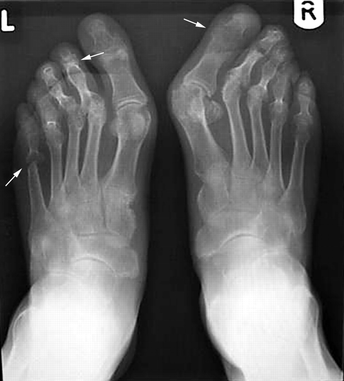
Radiograph of the feet of a patient with symmetrical polyarthritis with ulnar deviation of the hands and fibular deviation of the feet. The radiograph reveals joint fusion at multiple sites (arrows on top) and the fifth left metatarsal phalangeal head and adjacent proximal phalanx demonstrate pencil in cup deformity (arrow on left), features which help distinguish this as psoriatic arthritis rather than rheumatoid arthritis (RA). This case illustrates how imaging has the potential to clarify that the diagnosis is what clinically masquerades as RA.
A hypothesis in PsA is that enthesitis arises at sites of high shear and compression forces, with the additive interaction between mechanical stress, microtrauma, tissue repair mechanisms, and bacterial molecules variably leading to inflammation.19 PsA could represent a disease where the local microenvironment response to tissue stresses partly dictates the clinical expression of disease. This model also suggests a possible explanation for the observation that PsA, on occasion, may be associated with a preceding joint injury.20,21 Further, there may be a link to the skin component as injury to the skin is known to be a factor in expression of psoriasis. A curiosity is why psoriasis does not happen with every injury and why injury to joints of the patient with PsA associates even less frequently.
THE BONE IN PSA
From a historical perspective, bone scintigraphy showed that osteitis is a prominent feature of PsA but not RA.22,23 This is also seen in current MRI imaging in PsA. The basis for this is incompletely understood but seems to be linked to the close functional interdependence between the enthesis, the more broadly defined “functional” enthesis, and adjacent bone.5
CONCLUSIONS
Unlike RA, in which joint disease has a microanatomical basis centred on the synovium, imaging studies suggest that much of PsA pathophysiology is centred on the enthesis organ. This concept provides a unifying basis for a number of features of PsA, including disease symptoms, localisation, and imaging findings.
Acknowledgments
To all my colleagues in Leeds who make this work possible and to Prof M Benjamin in Cardiff for our ongoing fruitful collaboration on enthesis.
REFERENCES
Footnotes
-
D McGonagle is funded by the Medical Research Council, UK