Article Text

Abstract
Objective: There is evidence that atherosclerosis may contribute to the initiation or progression of osteoarthritis. To test this hypothesis, the presence and severity of hand osteoarthritis (HOA) was compared with markers of atherosclerotic vascular disease in an elderly population.
Patients and Methods: The AGES Reykjavik Study is a population-based multidisciplinary study of ageing in the elderly population of Reykjavik. In a study of 2264 men (mean age 76 years; SD 6) and 3078 women (mean age 76 years; SD 6) the severity of HOA, scored from photographs, was compared with measures of atherosclerosis. These included carotid intimal thickness and plaque severity, coronary calcifications (CAC) and aortic calcifications and reported cardiac and cerebrovascular events.
Results: After adjustment for confounders, both carotid plaque severity and CAC were significantly associated with HOA in women, with an odds ratio of 1.42 (95% CI 1.14 to 1.76, p = 0.002) for having CAC and 1.25 (95% CI 1.04 to 1.49, p = 0.016) for having moderate or severe carotid plaques. Both carotid plaques and CAC also exhibited significant linear trends in relation to HOA severity in women in the whole AGES Reykjavik cohort (p<0.001 and p = 0.027, respectively, for trend). No significant associations were seen in men. Despite this evidence of increased atherosclerosis, women with HOA did not report proportionally more previous cardiovascular or cerebrovascular events.
Conclusions: The results indicate a linear association between the severity of HOA and atherosclerosis in older women. The pathological process of HOA seems to have some components in common with atherosclerosis. Prospective studies may help elucidate the possible mechanisms of this relationship.
Statistics from Altmetric.com
Osteoarthritis is the most common of the musculoskeletal diseases, causing a huge burden of morbidity and disability, particularly in the elderly. The disease is manifest in all joint structures: cartilage, subchondral bone, synovium, capsule and ligaments. In many cases, there is also evidence of a generalised disorder with a symmetric polyarticular affection, most evident in hand osteoarthritis (HOA).1 2 The mechanisms controlling the initial events leading to osteoarthritis in a joint and the subsequent progression of the disease are mostly unknown.
Several investigators have found evidence implicating the vascular system in the development or progression of osteoarthritis.3 A number of studies has shown an association between osteoarthritis and cardiovascular comorbidity4 5 and even cardiovascular deaths.6 7 Others have described both venous and arterial pathology in the subchondral bone that apparently contribute to the progression of osteoarthritis.8 9 With the advent of magnetic resonance imaging, there is mounting evidence that the subchondral bone is directly involved in the development and progression of osteoarthritis.10 Ghosh and Cheras11 have also described a more systemic dysregulation of coagulation and fibrinolysis in osteoarthritis in both animal and human studies, and heparin-related drugs have had some success in the treatment of osteoarthritis.12 A recent report has also suggested that increased arterial stiffness may be a feature of HOA.13
The AGES Reykjavik study is a population-based study of elderly Icelanders, aged 66 years and older.14 Extensive information regarding the cardiovascular system has been gathered as well as high quality photographs of the hands. In this study we examined the relationship between photographic HOA and vascular pathology and its risk factors.15
Patients and methods
The AGES Reykjavik study is a population-based study of 5764 elderly individuals from the 40-year-long Reykjavik study. They were aged between 66 and 96 years and were randomly recruited between 2002 and 2005. Details of the investigations are described in the study’s baseline article.14 The participants underwent extensive laboratory and imaging investigations including high quality hand photographs.
Measures of atherosclerosis
The atherosclerotic outcome parameters were based on common carotid intima-media thickness (CIMT) and carotid plaque severity measured on ultrasound images and calcium in the coronary arteries and entire thoracic aorta measured on computed tomography (CT) images. Ultrasound images were acquired with a Sequoia C256 (Siemen Medical Systems, Erlangen, Germany). Standard B-mode images of the CIMT were acquired for the predefined segment of each common carotid artery (CCA; right and left) at defined interrogation angles using Meijers arc. Standard images were obtained from four angles at each site. The mean CIMT of the near and far walls were determined from a single image at each interrogation angle for both the right and left CCA. The average of all these CIMT values comprised the CIMT outcome parameter. The intima-media thickness analysis protocol is based on a technique described in detail elsewhere.16 The presence of atherosclerotic lesions of the left and right carotid bifurcation and internal carotid artery (Carotid plaques) was quantified on line during the ultrasound examination. The most severe lesions per segment were assessed in a semiquantitative manner as none, minimal, moderate and severe lesions, as described by Van der Meer et al.17 Images for calcium scoring were acquired using a Siemen Somatom Sensation 4 multi-detector CT (Siemen Medical Solutions, Erlangen, Germany) with prospective ECG triggering. The entire heart and thoracic aorta was scanned sequentially in two separate acqusitions in the cranio-caudal direction during suspended inspiration. Calcium in the coronary arteries and the thoracic aorta was quantified using the Agatston method. Coronary calcification (CAC) was expressed as a sum score for all four coronary arteries and the calcium in the thoracic artery was expressed as a sum score for the ascending aorta, descending aorta and the aortic arch. Interobserver variability assessment showed high average correlation between the five readers and an expert Multi-Ethnic Study of Atherosclerosis (MESA) reader (r = 0.94) based on the re-analysis of 200 scans. The CAC analysis technique is described in more detail elsewhere.18
Questionnaire information regarding cerebral stroke and cardiac events (myocardial infarctions, percutaneous coronary intervention or coronary artery bypass grafting) was used with regard to previous vascular events.
Hand photographs
The photographs were taken with a Fuji FinePix 6800 Zoom camera taken at 2800 × 2200 pixels. The camera was mounted on a tripod with a fixed distance to a velvet board with markers for thumb positioning. Photographs were available for 2191 participants of the initial 2300 (924 men and 1267 women) and 3151 of the remainder (1340 men and 1811 women). A scoring system for the reading of photographic HOA has been developed by two of the authors (HJ, GPH). With the help of a reference photo collection, it has been shown to have adequate reproducibility.19 The global estimates for the three joint sites (distal interphalangeal, proximal interphalangeal and carpometacarpal 1) used in this study were based on this scoring system, but with the added requirements of bilateral involvement of digits 2 or 3 for the distal interphalangeal and proximal interphalangeal joints. Definite nodal osteoarthritis (score 2 or higher) on one side, or bilateral suspected osteoarthritis (scores of 1) were thus classified as 1 (some evidence of HOA). Bilateral definite nodal osteoarthritis was required for a score of 2 (definite HOA) and bilateral definite osteoarthritis plus one or more severely affected joints were required for a global score of 3 (severe HOA) at each site. An aggregate severity score of 0–9 was thus constructed. On analysis, this aggregate score was subsequently truncated at 0, 1, 2, 3 and 4 or more (4+) for statistical purposes.
The reference photo collection is available on the IHA home page: http://www.hjarta.is/utgafa/hand-photography-as-a-method-for-diagnosing-hand-osteoarthritis.
Statistics
Results from the first 2300 participants constituted a predetermined dataset for the purposes of initial analysis, ie, an exploratory sample. Mean age at entry was 76 years (SD 6) for both genders. For subsequent validation of relevant findings, the remainder of the AGES Reykjavik study group (n = 3464), which has a similar gender and age distribution and a comparable prevalence of HOA, was used. Finally, analysis was done on the total sample.
HOA was used as a predictor for cardiovascular outcomes measures in two ways. First, as a binary predictor: HOA present versus HOA absent; then, as a five-point severity score (0–4). Carotid plaque was considered as a binary outcome variable in terms of present or absent. CAC was both considered as a binary outcome variable in terms of present or absent and as a continuous outcome analysed on the scale ln (CAC + 1) assuming normally distributed errors. Their association as binary outcomes was analysed separately for men and women using logistic regression models, with osteoarthritis severity as a binary predictor variable and as an ordinal predictor variable (0–4). The analysis was performed with and without account for confounding of age and smoking history (three-level variable: never, previous, current). Other possible confounders of the association were investigated: body mass index (BMI), hypertension, pulse pressure, serum cholesterol, low-density lipoprotein, triglycerides, high sensitivity C-reactive protein (hsCRP) and the use of statins.15 These additional variables did not have an effect on the interpretation of the associations presented. Similar analysis for CAC as a continuous outcome was performed with a linear regression model. Test of trend was computed using standard orthogonal linear contrasts appropriate for the five-point severity scale using the weights −2, −1, 0, 1, 2. Adjusted means or proportions were adjusted to mean levels of the covariates adjusted for. The statistical analyses were performed using SAS/STAT software version 9.1 and R software version 2.5.0.
Results
The prevalence and severity of HOA was higher in women than men, and women therefore accounted for a greater proportion of those with higher HOA scores. The prevalences are shown in table 1. The HOA severity scores correlated positively with age (Rs = 0.13, p<0.001) and negatively with smoking in both genders (Rs = −0.11, p<0.001).
Prevalence of aggregate HOA scores in the study population
Analysis of the initial sample of 2300, the subsequent validation sample and the total sample are shown in table 2. On uncorrected bivariate analysis, three vascular variables: carotid plaques, CAC and aortic calcifications showed significant correlations with both the presence and the severity of HOA in women. Age and smoking affected this relationship differently as both HOA and vascular pathology increased with age, whereas smoking was associated with increased vascular pathology but less photographic HOA. After adjustment for age, smoking, cholesterol, triglycerides, BMI, pulse pressure and statin use in the whole sample, both carotid plaques (odds ratio (OR) 1.25, 95% CI 1.04 to 1.49, p = 0.016) and CAC (OR 1.42, 95% CI 1.14 to 1.76, p = 0.002) showed a significant association with the presence of HOA. (table 2).
Analysis of the vascular variables in relation to the presence or absence of HOA
The prevalence of previous cardiac events in the study group was 8.8% in women without HOA and 7.4% in those with HOA (p = 0.039). Similar figures for men were 26.9% and 24.7%, respectively (p = 0.33). Obviously, this finding was marginal due to the low prevalence in women and was not confirmed in the validation group. No significant associations or linear trends were seen in men.
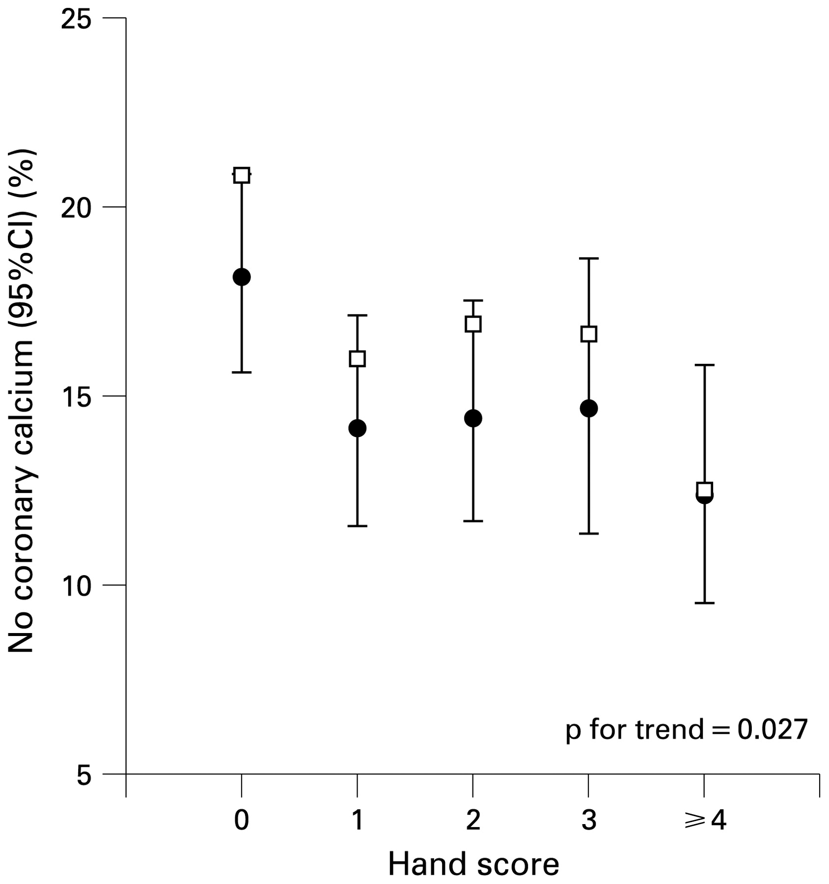
Both carotid plaques and CAC showed clear evidence of a linear relationship with HOA severity in women. The proportion of women in the AGES Reykjavik study with moderate or severe carotid plaque thus increased with the severity of HOA, with a linear trend significance of p<0.001 (fig 1). In the case of CAC, the proportion of patients with no detectable CAC decreased with increased severity of HOA (p = 0.027 for trend; fig 2).

Percentage of women (n = 3078) with carotid plaque score of moderate or higher in relation to hand osteoarthritis severity. The results (estimate, 95% CI) are adjusted for age and smoking to the profile of the reference group. Rectangles represent unadjusted values.
Percentage of women (n = 3078) with no coronary calcifications in relation to hand osteoarthritis severity. The results (estimate, 95% CI) are adjusted for age and smoking to the profile of the reference group. Rectangles represent unadjusted values.
Discussion
We examined the relationship between HOA and atherosclerosis in a population-based study of 3078 women and 2264 men. The female group showed a positive association between HOA and atherosclerosis in the coronary and carotid arteries, measured by two different imaging methods. These results remained significant when adjusted for age and other potential confounders, and were seen in both the initial sample as well as in the validation sample suggesting a true relationship. This relationship was linear, with increasing vascular pathology with HOA severity. Men showed no significant associations between HOA and vascular pathology. There was no evidence of an increase in cerebrovascular or cardiovascular events in the HOA group for either men or women.
Our results lend support to theories indicating that vascular pathology is an integral part of the osteoarthritis process, possibly contributing to the initiation or progression of HOA. In their article on vascular mechanisms in osteoarthritis, Ghosh and Cheras11 suggested a pathway in which osteoarthritis synovitis leads to a state of hypercoagulation and hypofibrinolysis, and subsequently to circulatory disturbances in the subchondral bone contributing to the perpetuation of cartilage destruction and the pathophysiological process of osteoarthritis. Areas of bone marrow oedema in the subchondral bone detected by magnetic resonance imaging are strongly associated with the symptoms and progression of osteoarthritis.10 Histologically, bone marrow oedema is areas of necrosis and remodelling20 and may well represent ischaemic areas in bone, at least in some cases. It has been postulated that subchondral bone may lead to cartilage damage through mechanical changes in the bone, and recently a more direct pathway by which bone-derived cytokines mediate degradation of cartilage has been identified.21
Inflammatory diseases such as rheumatoid arthritis are associated with accelerated atherosclerosis and increased cardiovascular mortality. This has been attributed to many factors, including impaired endothelial function, less favourable cardiovascular risk factor profiles and inflammation characterised by increased serum hsCRP.22 Inflammation in osteoarthritis is much less marked, but synovitis is often present and serum hsCRP levels are increased in active HOA.23 In this study, we found no association between study hsCRP levels and HOA, nor between hsCRP and the vascular changes related to HOA in this elderly study group (data not shown).
Other potential explanations of the relationship between HOA and atherosclerosis must also be considered. In view of the strong genetic inheritance of HOA, genetic factors can contribute to this relationship.24 25 The KLOTHO gene, which codes for an anti-ageing protein has recently been implicated both as a susceptibility factor for HOA in women26 and as a candidate gene for atherosclerosis.27
The slight inverse association between HOA and cardiovascular events despite increased vascular pathology and strong positive associations between the vascular variables and events was an unexpected finding that could not be positively confirmed in the control group. In a cross-sectional study of this kind, the explanations are not clear, but one possible mechanism relates to the intake of polyunsaturated fatty acids. In Iceland, cod liver oil or omega-3 fatty acids are widely recommended for symptomatic osteoarthritis, and the intake of these substances was significantly more frequent in the HOA group (data not shown). Hypothetically, a marginal protective effect on cardiovascular events and survival by long-term use of these substances could contribute to our current findings, ie, a low prevalence of cardiac events in those using them despite vascular disease.28
Our observed differences between men and women may be explained by a number of factors. The epidemiology of HOA in men and women is quite different, with HOA being more common in men until middle age, but after that more prevalent in women. We did not find evidence to suggest that symptomatic HOA was associated with more atherosclerosis (data not shown), but it is also several times more common in women, suggesting the possibility of different disease mechanisms.29 Add that to a possible mortality effect of HOA in men6 and the fact that men of this age have more atherosclerosis and higher mortality, and these factors may obscure a relationship of this kind. Furthermore, photography is a less sensitive method for the diagnosis of HOA in men than in women and compares less well with radiographic osteoarthritis.29
The main weakness of our findings is related to the study’s cross-sectional nature, limiting conclusions regarding a possible cause and effect relationship between HOA and vascular pathology. Our method of diagnosing HOA is not necessarily comparable with previous studies using radiography, but there may be advantages in using photography, identifying visible nodes. We had an unusually strong negative correlation between HOA and smoking compared with radiological studies and, compared with radiography, we may have identified a subset of HOA in which systemic and/or vascular factors are particularly relevant. In an ancillary study in which we had clinical examination and radiograph scores in 381 subjects29 there were indications that the clinical examination scores may have a stronger association with atherosclerosis than radiographic scores (data not shown).
Despite reports of vascular disturbances in osteoarthritis, this is the first report that confirms increased atherosclerosis in women with HOA. The exact mechanisms cannot be determined from this cross-sectional study, but the presence of HOA may have to be accounted for in future studies of atherosclerosis risk factors. A prospective part of the AGES Reykjavik study is now under way, and we hope that the acquisition of longitudinal data will help us to clarify some of the relevant mechanisms. Other research questions of particular interest are whether the relationship between HOA and atherosclerosis observed in this study is present in osteoarthritis at other sites such as the knee and hip, and whether the course of osteoarthritis can be affected by treatment directed at vascular pathways.
REFERENCES
Footnotes
Funding This study has been funded by National Institutes of Health contract N01-AG-12100, the NIA Intramural Research Program, Hjartavernd (the Icelandic Heart Association) and the Althingi (the Icelandic Parliament). The study was also supported by the Icelandic Osteoarthritis Fund and the University of Iceland Science Fund.
Competing interests None.
Ethics approval The study was approved by the Icelandic National Bioethics Committee, VSN 00-063, the Icelandic Data Protection Authority and the Institutional Review Board serving NIA.