Article Text

Statistics from Altmetric.com
Decreased hand function is a major contributor to disease burden in rheumatoid arthritis (RA) and is present in most patients with RA at diagnosis.1 2 Recent research showed that hand function is reduced in the symptomatic prearthritis phase of clinically suspect arthralgia (CSA) and is a reflection of subclinical tenosynovitis.3 Grip strength (GS) measured with a dynamometer had the highest sensitivity for decreased hand function and underlying tenosynovitis, compared with other assessment methods of hand function.3 Although this may suggest that the dynamometer could be a practical assessment in CSA to objectify functional impairments originating from subclinical joint inflammation, it needs to be determined if dynamometer-based GS assessments in CSA are sensitive to change and mirror the disease course of CSA. To our best knowledge, longitudinal studies on GS in the phases preceding RA diagnosis are lacking. We hypothesised that GS follows distinct natural trajectories in patients with CSA who have contrasting disease courses (RA development, persistent CSA symptoms without RA development and spontaneous resolution of arthralgia). Second, since it was recently shown that a temporary methotrexate treatment in CSA resulted in sustained improvements of subclinical joint inflammation, we hypothesised that GS is responsive to treatment in the CSA phase.4
Of the 117 patients in the placebo group, 21 patients developed RA; 35 patients achieved spontaneous resolution of pain; and 61 patients had persistent symptoms. Patients with CSA who progressed to RA were more often positive for anti-citrullinated protein antibodies (ACPA-positive): 52% vs 13% and 11% in patients with persistent and resolving complaints, respectively, and had a higher median MRI-detected inflammation score on baseline: 5.5 vs 4 in the other two subgroups. The subgroup of patients achieving resolution had somewhat less pain on inclusion: a median pain score of 40 (vs 50 in the other subgroups) and tender joint count (TJC) of 2 compared with TJCs of 3 and 4 in patients who progressed or had persistent complaints, respectively (online supplemental table 1). At trial inclusion, mean GS was 31.4 (2.3) in patients achieving resolution, 28.8 (1.7) in patients with persistent symptoms and 31.7 (3.2) in patients who later developed RA (online supplemental figure 1). Patients with subclinical joint inflammation on MRI and subclinical tenosynovitis in particular had lower GS: per point increase in tenosynovitis, GS decreased with −2.63 kg (95% CI −2.26 to −0.33).
Supplemental material
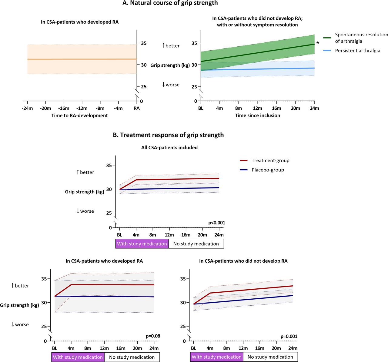
Studying the natural course of GS over time in the three patient groups revealed that GS remained stable in patients with CSA who did develop RA (−0.03 kg/month; 95% CI −0.26 to 0.19, p=0.76) or had persistent CSA complaints (0.02 kg/month; 95% CI −0.06 to 0.11, p=0.64). In patients who achieved pain resolution, GS increased with 0.16 kg/month (95% CI 0.06 to 0.27, p=0.002) (figure 1A). Thus, patients with resolving symptoms had improvement of GS, in contrast to patients who developed RA or had persistent CSA complaints. In support of these observations, unmodelled data and analysis of treatment response using time as a categorical variable are depicted in online supplemental figures 1 and 2, respectively. Hence, GS followed distinct natural trajectories in patients with CSA with contrasting disease courses.

The natural course of GS in CSA (A) and improvement on a temporary treatment in CSA (B). (A) Of the 117 patients in the placebo group, 21 developed RA; 35 achieved spontaneous resolution of pain; and 61 had persistent CSA symptoms. (B) GS of patients with CSA in the treatment group (n=119) was compared with that of the placebo group (n=117). Within the treatment group, 23 patients with CSA developed RA and 96 did not; in the placebo group, these numbers were 21 and 96, respectively. The bands represent the 95% CI of the estimated mean. *P=0.002. CSA, clinically suspect arthralgia; GS, grip strength; RA, rheumatoid arthritis.
We then studied whether GS is responsive to treatment in the CSA phase by comparing the treatment and placebo arm. Treatment induced a mean GS improvement of 1.97 kg over 2 years (95% CI 0.86 to 3.07, p<0.001), which sustained after treatment stop. Treatment-related improvements were present both in patients with CSA who developed RA (total of 44 participants, 23 in the treatment-group) (+2.47 kg; 95% CI −0.29 to 5.24, p=0.08) and in patients with CSA who did not develop RA (+2.04 kg; 95% CI 0.83 to 3.24, p=0.001) (figure 1B). Sensitivity analysis with GS of the weakest hand showed comparable results, except for a small spontaneous increase in placebo patients with persistent CSA complaints (+0.08 kg/month; 95% CI 0.003 to 0.16, p=0.04) (online supplemental table 2).
This study provides the first evidence that GS assessment is sensitive to change in patients with CSA with subclinical joint inflammation. While GS was reduced in CSA and remained so during progression to RA, it improved in patients with CSA with spontaneous resolution. Moreover, it also improved on treatment.
The observed treatment effect is in line with reported findings of sustained improvements in subclinical joint inflammation and patient-reported outcomes.4 The 2 kg improvement in GS is clinically relevant and quite comparable to reported improvements during the first year of treatment after RA diagnosis (around 3.5 kg).2 5 Hence, this study underlines that a temporary treatment in the CSA phase could improve hand function in the CSA phase and also in patients with CSA with subclinical inflammation who will not progress to RA.
Once tools for monitoring of disease activity in the CSA phase are developed, GS could be of value as a component of a multidimensional/composite score, as it supports the ‘sensitivity-to-change’ and ‘longitudinal construct validity’ items in the Outcome Measurement in Rheumatology (OMERACT) filter for instrument selection.6
In conclusion, GS is easily assessed in practice and responds to treatment in CSA, and its course could be of value for monitoring disease activity in the at-risk phase of CSA.
Ethics statements
Patient consent for publication
Ethics approval
This study involves human participants and was approved by the medical ethical committee of the Leiden University Medical Centre. The participants gave informed consent to participate in the study before taking part.
Supplementary materials
Supplementary Data
This web only file has been produced by the BMJ Publishing Group from an electronic file supplied by the author(s) and has not been edited for content.
Footnotes
Contributors DIK and AvdHvM designed the study. DIK, FW and EN collected the data. DIK analysed the data. All authors interpreted the data and wrote the report. AvdHvM was the principal investigator. All authors approved the final version of the manuscript.
Funding This work was supported by a ZonMW grant (Programma Translationeel Onderzoek), by the European Research Council under the European Union’s Horizon 2020 research and innovation programme (starting grant, agreement number 714312) and the Dutch Arthritis Society.
Competing interests None declared.
Provenance and peer review Not commissioned; externally peer reviewed.
Supplemental material This content has been supplied by the author(s). It has not been vetted by BMJ Publishing Group Limited (BMJ) and may not have been peer-reviewed. Any opinions or recommendations discussed are solely those of the author(s) and are not endorsed by BMJ. BMJ disclaims all liability and responsibility arising from any reliance placed on the content. Where the content includes any translated material, BMJ does not warrant the accuracy and reliability of the translations (including but not limited to local regulations, clinical guidelines, terminology, drug names and drug dosages), and is not responsible for any error and/or omissions arising from translation and adaptation or otherwise.